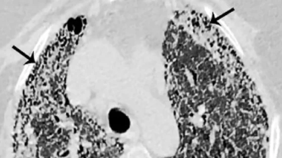
Representative non-contrast CT slices for two patients (left), with super-imposed segmentations (right). One artificial intelligence (AI) model was used to segment a cardiac mask (magenta line) and coronary artery calcium (red). A second AI model segmented left ventricular myocardium (purple), left atrial (green), left ventricle (light red), right ventricle (blue) and right atrial (yellow) volumes. Images/caption courtesy of Slomka et al. and Nature Communications.


![Advanced artificial intelligence (AI) models can evaluate cardiovascular risk in routine chest CT scans without contrast, according to new research published in Nature Communications.[1] In fact, the authors noted, the AI approach may be more effective at identifying issues than relying on guidance from radiologists. Representative non-contrast CT slices for two patients (left), with super-imposed segmentations (right). One artificial intelligence (AI) model was used to segment a cardiac mask.](/sites/default/files/styles/top_stories/public/2024-04/screen_shot_2024-04-23_at_10.44.32_am.png.webp?itok=O8wgPFEZ)