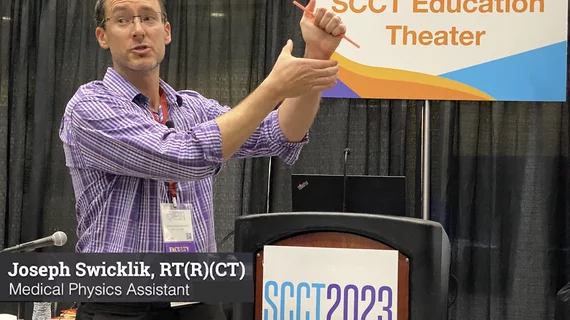
Joseph Swicklik, RT(R)(CT), Mayo Clinic, says magic tricks can help calm pediatric patients to overcome anxiety and make medical imaging exams run much smoother.
The company officially launched its Cleerly ISCHEMIA software for delivering noninvasive evaluations of CCTA exams in early 2024. This update provides a closer look at the software from a billing perspective.
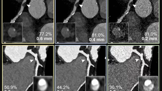
Examples of photon-counting coronary angiography showing how clarity improves as the thickness of the image is reduced. Top: 60-year-old female, with noncalcified plaque (arrowheads) and coronary stenosis (inset images). The reduced section thickness did not affect assessment in this patient. Bottom: 56-year-old female with calcified plaque (arrowheads) and coronary stenosis. The reduced section thickness leads to less calcium blooming and therefore a less severe percentage of stenosis. Photos courtesy of RSNA.
After a photon-counting CT, 54% of patients had their coronary artery disease classification downgraded.