A new three-dimensional (3D) MRI technique can measure strain in the heart without using potentially damaging gadolinium, according to new research out of the University of Warwick in the U.K.
The method, described in an Aug. 28 study published by Scientific Reports, uses a novel calculation technique to determine which heart muscles are not functioning properly and can do so without the threat of gadolinium affecting a patient’s kidney function.
"Using 3D MRI computing technique we can see in more depth what is happening to the heart, more precisely to each heart muscles, and diagnose any issues such as remodeling of heart that causes heart failure,” said Mark Williams, from Warwick Manufacturing Group (WMG), part of the University of Warwick, in a news release. “The new method avoids the risk of damaging the kidney opposite to what traditional methods do by using gadolinium."
This gadolinium contrast reacts with the magnetic field to create an image showing where dead muscle is in the heart and, hopefully, a subsequent diagnosis.
However, as more research comes out, the use of gadolinium is being associated with negative effects on the body, specifically the kidneys. This new method, the researchers say, can help avoid that and alleviate some of the stress that comes with undergoing an MRI.
"This new MRI technique also takes away stress from the patient, as during an MRI the patient must be very still in a very enclosed environment meaning some people suffer from claustrophobia and have to stop the scan, often when they do this they have to administer another dose of the damaging gadolinium and start again,” said Jayendra Bhalodiya, also with WMG Warwick. “This technique doesn't require a dosage of anything, as it tracks the heart naturally."
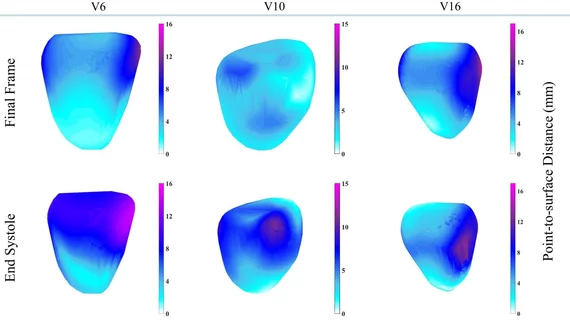
The researchers tested their hierarchical template matching model in an anonymized and publicly available dataset. They found 3D myocardial tracking could be done with 1.49 millimeter median error and a reduced maximum error of tracking by up to 24% compared to “benchmark” methods.
“It should be noted that our contribution is not limited to only cardiac 3D MRI analysis,” the team concluded. “The transformation function, which is extended in this article for 3D, can be used with other application areas where 3D imaging has already established standards.”

