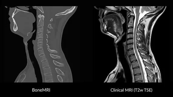
MRIguidance, a spin-off company of the University Medical Center Utrecht in the Netherlands, is developing an advanced MRI software that can characterize both soft tissue and bone without the use of radiation, according to a company news release published Oct. 16.
The anticipated software solution called BoneMRI will create three-dimensional (3D) CT-like images of human bone in a single imaging exam with conventional MRI scanners—which up until now have mainly been used to analyze soft tissue.
“Like all radiologists, I have been trained with the idea that MRI is mainly suitable for soft tissue imaging and x-ray based techniques are required to visualize bone,” Peter Seevinck, co-founder of MRIguidance and inventor of BoneMRI, said in the release. “But during my work as a scientist in the UMC Utrecht, I was getting more and more convinced that MRI data, when acquired in the right way, contains all the information necessary to also visualize bone.”
BoneMRI is being developed and clinically validated at several large hospitals in the Netherlands and Belgium and can be implemented into clinical workflow without additional desktop applications.
Recently, MRIguidance secured a €1.5 million ($1.9 million) investment from health-tech investors for the development and the market roll-out of the software, according to the release.
“The investment gives MRIguidance a great boost and will accelerate our efforts to bring BoneMRI to the patient,” said Roel Raatgever, CEO of MRI guidance. We are very happy to get a strong investors team on board.”

