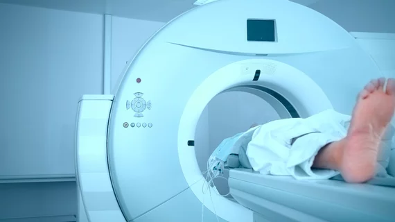
Notably lower T1 relaxation times, or how brain signals can weaken over time, may point to the presence of gadolinium in the brains of patients who’ve had more than one contrast-enhanced MRI exam, according to research published online Jan. 1 in Radiology.
A group of German researchers found no difference in MRI signal intensity in patients who received at least five doses of the macrocyclic gadolinium-based contrast agent (GBCA) gadobutrol—specifically Gadavist—and in controls with no exposure to the contrast agent. However, there was a significant difference seen in T1 relaxation times in the brain region called the globus pallidus.
“As an alternative explanation, the shorter T1 times in the globus pallidus may result from the accumulation of other elements, such as iron...our study showed that age influences T1 times in most of the analyzed brain areas, highlighting the necessity of age control in clinical studies investigating gadolinium retention by using MRI,” wrote Marc Saake, MD, from the University Hospital Erlangen in Germany, and colleagues.
For the study, two groups of patients who received gadobutrol between 2007 and 2011 and two groups of healthy controls with no exposure to the GBCA contrast agent underwent unenhanced brain MRI. The protocol included T1-weighted spin-echo and T1 and T2 mapping to determine visual signal intensity changes, signal intensity ratios of the globus pallidus to the thalamus and the dentate nucleus to the pons, and T1 and T2 relaxation times.
Group one consisted of 76 participants who had at least five contrast agent-enhanced examinations and normal kidney function; 84 participants in group two had at least one examination and impaired renal function. A total of 25 healthy participants and 35 participants with impaired renal function without history of contrast agent exposure were included for comparison.
Study results showed no visual changes or differences in signal-intensity ratios or in T1 and T2 mapping, however group 1 who had normal renal function and multiple administrations of gadobutrol showed lower T1 relaxation times in the globus pallidus compared with group 3, the control subjects with normal renal function.
Overall, T1 relaxation times of the globus pallidus were 26.2 milliseconds shorter on average and inversely correlated with the number of prior gadobutrol doses, according to the researchers.
While the team is unsure of whether the T1 relaxation time changes indicate potentially small amounts of gadolinium in the globus pallidus, the overall research supports the idea that patients should be informed about the possibility of gadobutrol brain retention before undergoing contrast enhanced imaging MRI exams.
The study was supported by Bayer.
Related MRI Contrast Agent Safety Content:
A deep dive into gadolinium-based adverse reactions
Allergic reactions to iodinated CT contrast increase likelihood of sensitivity to GBCAs
Researchers detail data on gadolinium-related adverse reactions
Radiologists must take a data-driven approach to discuss gadolinium, mitigate liability risk
Radiologists see potential to reduce GBCA administration with new synthetic MRI technique
Gadolinium-based contrast agents are safe, even at higher doses, new research suggests
Gadolinium debate rages on, with radiologist questioning recent GBCA liability guidance
ACR committee proposes new term for symptoms associated with gadolinium exposure
Closing the knowledge gap on gadolinium retention risks
Radiologists find direct evidence linking gadolinium-based contrast agent to higher retention rates
AI software that eliminates need for gadolinium contrast during imaging exams wins patent
Radiology, other multispecialty groups urge caution with GBCAs during interventional pain procedures
Cardiac MRI contrast agents are low-risk and safe for ‘overwhelming’ majority of patients
Health orgs publish special report about gadolinium retention, GBCA use in imaging
Rodent brains retain gadolinium after repeated administration of GBCA a year after injection
Advanced MRI mapping spots traces of gadolinium in the brain invisible during conventional scanning
Radiologists should keep patients’ best interests in mind to mitigate gadolinium liability risk

A recent graduate from Dominican University (IL) with a bachelor’s in journalism, Melissa joined TriMed’s Chicago team in 2017 covering all aspects of health imaging. She’s a fan of singing and playing guitar, elephants, a good cup of tea, and her golden retriever Cooper.