
Virtual imaging trials can be used to safely test and optimize CT and radiography techniques for more reliable pandemic-related care, researchers reported recently.
Doctors from Duke University described their experience combining computational models of patients with the novel virus with COVID-19 imaging simulators Friday in AJR. The virtual framework allows users to test how different modalities can be used to battle the pandemic, which is difficult to do in real time when healthcare workers are focused on treating patients.
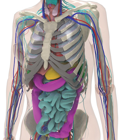
“In virtual imaging trials (also known as virtual clinical trials or in silico trials), the imaging experiment is emulated using computational models of patients combined with validated imaging simulators,” Ehsan Abadi, PhD, with Duke’s radiology department, and colleagues wrote. “Virtual imaging trials are highly advantageous in that the same subjects can be imaged with different scanners and settings, and the exact truth of the anatomic and physiologic features of the subjects are a priori known.”
The team created computational patients using a 4D extended cardiac-torso model, which is based on clinical data and includes “thousands” of anatomic structures. Abnormalities associated with COVID-19 were taken from 20 CT scans of patients with the virus and incorporated into the human phantom model.
In total, Abadi et al. developed three different simulation models, which could be imaged using either a CT or radiography simulation tool developed at the Durham, North Carolina, university.
Based on subjective feedback, the simulated abnormalities looked “realistic” in shape and texture, the authors noted, offering a foundation for use in virtual imaging trials.
The authors said they hope to use these trials to compare radiography and CT methods and define COVID-specific protocols for improved screening and follow-up in patients with the disease. That also includes optimizing radiation dose when imaging patients with the coronavirus.
Such attempts may also prove useful for training artificial intelligence approaches.
“Because virtual imaging trials have predefined models of the disease, they can provide a valuable tool to train and evaluate either principle-informed or machine learning algorithms designed for disease classification and quantification,” the authors concluded.
Read the entire study in the American Journal of Roentgenology.

Matt joined Chicago’s TriMed team in 2018 covering all areas of health imaging after two years reporting on the hospital field. He holds a bachelor’s in English from UIC, and enjoys a good cup of coffee and an interesting documentary.