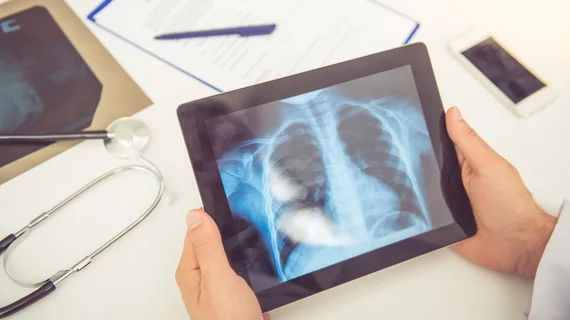
Radiologists more accurately spotted major abnormalities on chest X-rays with the help of a deep learning-based detection system, according to results of a new randomized study.
A number of prior investigations have revealed the power of artificial intelligence algorithms as a second reader, yet all past research has asked rads to interpret exams sequentially, experts explained Tuesday in Radiology. Looking at images with CAD assistance and then without quickly introduces reader order bias, they noted.
Utilizing a randomized, crossover design, the investigators showed imaging professionals—including an experienced thoracic radiologist— were quicker and more accurate at detecting abnormalities when using CAD.
“In the rigorous setting of our study, which minimized bias, the diagnostic performance of the radiologists was substantially improved with the assistance of the DLD system,” Jinkyeong Sung, and colleagues with Asan Medical Center’s Department of Radiology in Seoul, South Korea, wrote on March 23.
To arrive at their conclusions, the authors retrospectively collected 114 abnormal and 114 normal chest radiographs between January 2016 and December 2017. Six readers interpreted each image using the deep learning system and then again without it, abiding by the crossover design and washout period.
With the detection software, radiologists improved in a number of areas. For instance, localization rose from 0.90 to 0.95 and area under the receiver operating curve scores jumped from 0.93 to 0.98. At the same time, per-lesion sensitivity and specificity both increased along with per-image sensitivity.
Furthermore, the deep learning-based system dropped read times from 10-65 seconds down to 6-27 seconds, the authors reported.
Sung and co-authors encouraged future studies to go beyond the single-center design utilized for their own research in order to make sure this system is ready for clinical use.
“Further studies in other institutions and countries are needed to ensure generalizability,” the authors concluded. “In addition, future studies in a real-world setting will help determine the usefulness of the DLD system.”

