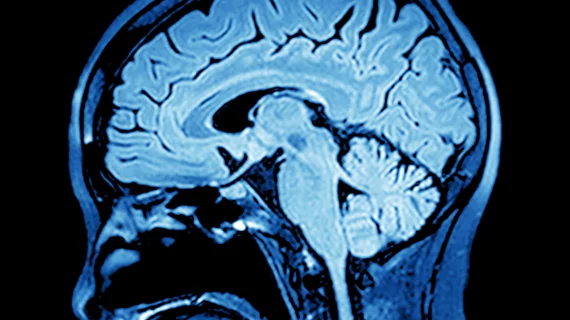
When imaging brain tumors such as gliomas, machine learning may advance the use of imaging and augment clinical care for patients, according to a review published Oct. 17 in the American Journal of Roentgenology—specifically in tumor segmentation and MRI radiomics.
Although there is great potential for improving clinical diagnostics, prognostics and decision-making with machine learning in glioma imaging, there are also numerous challenges that must be considered for successful implementation, wrote lead author Yvonne W. Lui, MD, a radiologist at New York University Langone Medical Center, and colleagues.
To overcome future challenges, researchers believe characterizing brain tumors through more stable, reproducible automated methods can help gather more quantitative data to better assess, define and characterize tumors.
Advances in convolutional neural network (CNN) deep learning architectures for tumor segmentation are contributing to the progress of MRI radiomics.
“Deep learning algorithms can be stacked to achieve end-to-end training from image to segmentation to classification to outcome prediction,” according to Lui et al. “These stacked modules, in fact, can be used to assist one another in training, potentially improving overall performance. Deep learning is capable of learning highly intricate and abstract patterns from multimodal imaging that may not be readily apparent to observers.”
However, multimodal imaging can lead to “overfitting”—when a machine learning algorithm is too specific to the training set and useless in other cases—particularly with deep learning approaches that require very large training datasets for accurate models.
“Such datasets are difficult to amass and curate, though there are efforts under way to pool data,” the researchers wrote. “Reliable methods of feature selection can reduce the number of free parameters and eliminate redundant or uninformative features, and there are multiple strategies for dimensionality reduction of the feature vector.”
Another way to address the issue of limited data in brain tumor radiomics is to use transfer learning, the researchers noted. Transfer learning is where the hierarchy of CNN features learned for a separate but related task is used and fine-tuned for the underpowered task of interest. This method allows the underpowered task-in-hand to borrow statistical power from the larger training dataset, while still being fine-tuned for the task-in-hand.
“Machine learning has often been criticized as being a black-box approach that does not inform regarding underlying mechanisms of disease,” according to the researches. “Recent work in visualization of deep learning models is, however, providing some avenues for opening the black-box and visualizing these biomarkers.”
Overall, if larger amounts of well annotated data are obtained, the researchers wrote, good generalization across data sets can be achieved.
“As more machine learning techniques are developed that address specific issues relevant to brain tumor segmentation and radiomics, the results almost certainly will improve,” the researchers wrote. “Incorporating information from complementary imaging modalities and clinical data are sure to broaden the relevance of such techniques. Moreover, as we gain better understanding of machine learning theory, choosing machine learning architecture most suitable for particular applications will contribute to advances.

A recent graduate from Dominican University (IL) with a bachelor’s in journalism, Melissa joined TriMed’s Chicago team in 2017 covering all aspects of health imaging. She’s a fan of singing and playing guitar, elephants, a good cup of tea, and her golden retriever Cooper.