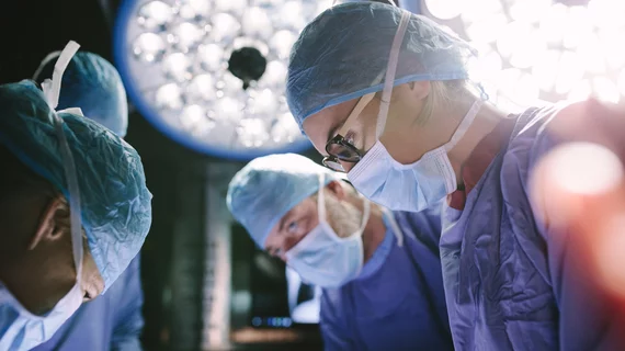
The University at Buffalo has received a national grant to further develop a medical imaging technique used during leg ulcer care, the college announced recently.
UB biomedical engineers and surgeons will use the $1.6 million National Institutes of Health award to study photoacoustic tomography and improve its ability to assess post-surgical blood circulation in those with chronic leg ulcers. Such injuries take time to heal and some never do, leading to costly treatments, limited mobility and a higher risk of death.
Researchers involved in the four-year project believe their imaging tool can overcome the limitations of MRI, ultrasound and X-rays for assessing blood perfusion after surgery.
“Our idea is, after the UBMD vascular surgery team has done the surgery to restore proper blood flow, we’ll do the imaging,” said Jun Xia, PhD, associate professor in the Department of Biomedical Engineering. “We’ll compare the results with the imaging acquired before the surgery to see if there is any change in the blood perfusion to the ulcer. If there is a positive change, then we know the surgery was successful,” Xia, who received the grant, added July 7.
Current practice requires surgeons to wait months before assessing whether blood flow has been restored. If blood cannot reach the injured tissue, cells will eventually die off and the injury turns into a chronic condition. Xia and co-investigators believe they can decrease that lag time to two weeks or less.
“This will allow surgeons to identify earlier than current imaging techniques whether the surgery was successful,” Xia added.

Matt joined Chicago’s TriMed team in 2018 covering all areas of health imaging after two years reporting on the hospital field. He holds a bachelor’s in English from UIC, and enjoys a good cup of coffee and an interesting documentary.