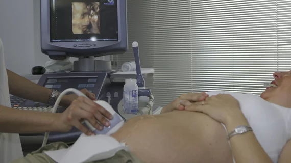
A new study offers insight into the prenatal development of children who are eventually diagnosed with autism spectrum disorder (ASD) by using ultrasound to identify early anatomical anomalies.
Many children on the spectrum are not diagnosed until several years into their lives, often during crucial developmental stages. Since most ASD diagnoses occur because of behavioral symptoms, the researchers note that there is a growing interest in tracking prenatal organ development in these children as a means for a more accurate and timely diagnosis.
“A growing body of evidence suggests that the initial signs of ASD emerge during early childhood and possibly even before birth,” corresponding author Professor Idan Menashe, a member of the Centre and the Department of Public Health in the Faculty of Health Sciences at Ben-Gurion University of the Negev in Israel, and coauthors explained. “Recent postnatal studies have found indications of the prenatal onset of abnormal neurodevelopment in children with ASD.”
The study, published in Brain,[1} retrospectively examined second trimester ultrasound features and long-term post-natal development of nearly 700 children, including 229 with ASD, 201 typically developing siblings (TDS) and 229 typically developing children from the general population (TDP).
Among the fetuses who later developed ASD, 30% displayed anomalies on their prenatal ultrasound, which was three times higher than the TDP group. UFAs of the urinary system, heart, head and brain were the most frequently associated with ASD diagnosis, and multiple abnormalities on the same exam were significantly linked with the neurodevelopmental disorder.
The researchers noted that the severity of the anomalies were also linked with patients’ reported ASD symptom severity.
“These UFAs, which can be detected in standard prenatal anatomy ultrasound surveys conducted during mid-gestation, could form the basis of new prenatal screening approaches for ASD,” the experts suggested. “The results of such prenatal screening will reveal fetuses at risk to develop ASD and may facilitate their earlier diagnosis, a factor that has already been shown to optimize the long-term outcomes of ASD treatment.”
The authors note that previous research has indicated that early diagnosis and treatment of ASD can increase social abilities by up to three times and understanding a child’s likelihood of developing ASD before birth could initiate therapy at the earliest possible time. They also acknowledged that these findings should be considered alongside many other factors, as autism is considered a spectrum disorder and although prenatal imaging could offer insight into the development of ASD, it should not be used as a solo diagnostic tool.
You can view the full study here.
Related Autism Content:
6 tips for making MRI exams more autism-friendly
MRI scans link atypical growth of key brain structure during infancy with autism
MRI features uncover differences between the brains of autistic girls and boys
Placental MRI can predict adverse pregnancy outcomes early on in gestation
Fetal structural anomalies to blame for many pregnancy terminations
Maternal social disadvantages linked with reduction in baby brain volumes at birth
Reference:

