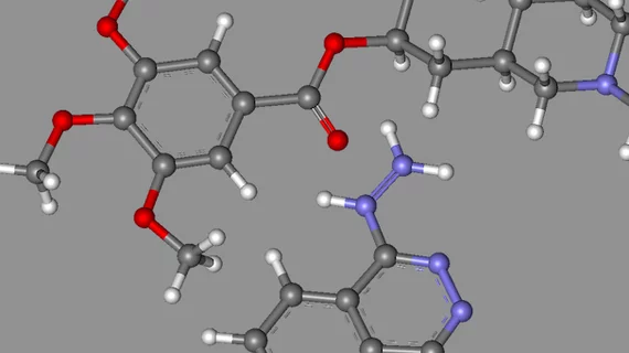
A team of University of California Los Angeles and California Institute of Technology in Pasadena researchers have developed a new molecular imaging technique that may speed up drug discovery and is being dubbed as “a new day for chemistry” by the scientific community.
The technique is able to map the structure of small organic molecules, such as pharmaceuticals and hormones, with electron diffraction imaging that is commonly used to chart larger proteins, according to a report published Oct. 19 by Science Magazine.
Until now, x-ray crystallography and nuclear magnetic resonance spectroscopy have been the sole options to determine chemical structures. Utilizing electron diffraction, the new technique sends an electron beam through a crystal and—as in x-ray crystallography—determines a molecules structure from diffraction patterns, according to the article.
For the study, researchers rotating the crystal and tracking how the diffraction pattern changed. They ultimately ended up getting an image that looked more like a molecular CT scan and enabled them to get structures from crystals one-billionth the size of those needed for x-ray crystallography. The research was published online Oct. 16 in ChemRxiv.
See Science Magazine’s entire article below.

