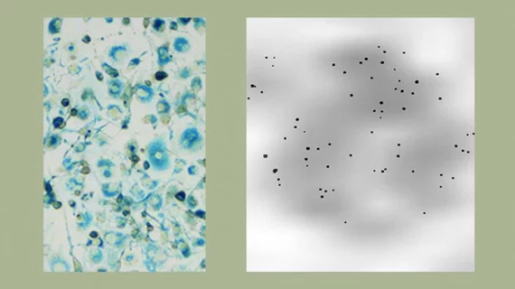
Researchers from the Keck School of Medicine at University of Southern California (USC) successfully used high-resolution MRI and a cell-labeling technique to visualize less than 100 cells, which may allow clinicians to assess the effectiveness of immune cell- and stem cell-based therapies to treat cancer.
The research was published in the July issue of the International Journal of Nanomedicine.
“The application of this new method is very broad,” said co-researcher Danny J.J. Wang, PhD, professor of neurology at the Mark and Mary Stevens Neuroimaging and Informatics Institute at USC, in a prepared statement. “We used standard clinical equipment and FDA-approved drugs in the hopes that the results can translate quickly from bench to bedside.”
Wang and colleagues tested the new method of labeling cells which allowed the cells to be easily seen in an MRI scanner.
The researchers labeled cell macrophages with three FDA-approved drugs—herapin, protamine and ferumoxytol—and then used MR to capture images of the macrophages. In some trials, Wang and his team were able to detect as few as 62 cells labeled with the drugs, according to the USC news release.

