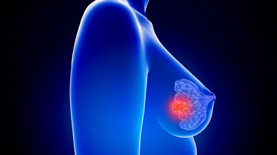
A hybrid nuclear imaging approach discovered multiple breast cancer biomarkers that may help physicians better diagnose the disease, according to a new study.
Researchers compared healthy tissue in the breasts of patients with malignant and benign tumors, noting that PET/MRI could visualize differences in biomarkers between each group. Such markers, they noted in the study to-be published in the January issue of the Journal of Nuclear Medicine, could be used in risk-adapted and reduction screening strategies.
“As hybrid PET/MRI scanners are increasingly being used in clinical practice, they can simultaneously assess and monitor multiple imaging biomarkers—including breast parenchymal uptake—which could consequently contribute to risk-adapted screening and guide risk-reduction strategies,” Doris Leithner, MD, research fellow at Memorial Sloan Kettering Cancer Center in New York, said in a statement.
Early detection of the disease remains central to a patient’s prognosis and chance of survival. Screening mammography has brought cancer mortality down by nearly 30%, but it does have sensitivity limitations, particularly in women with dense breast tissue. These “shortcomings,” as Leithner described them, necessitate “refinements” in modalities used in breast cancers screening, she said in the release.
The team included 141 patients who underwent combined 18F-FDG PET/MRI and registered imaging abnormalities on their mammogram or sonogram (considered BI-RADS 4 or 5). Each patient had several imaging biomarkers found in the tumor-free breast, opposite of the abnormality. Those included: background parenchymal enhancement and fibroglandular tissue from MRI, mean apparent diffusion coefficient and breast parenchymal uptake from 18F-FDG PET.
Two independent experts assessed 100 malignant and 41 benign lesions. In the tumor-free breast tissue, background parenchymal enhancement and breast parenchymal uptake dropped and varied greatly between women with benign and malignant lesions, the analysis found. The other two biomarkers, however, did not show any significant difference between the groups.
“Based on these results, tracer uptake of normal breast parenchyma in 18F-FDG PET might serve as another important, easily quantifiable imaging biomarker in breast cancer, similar to breast density in mammography and background parenchymal enhancement in MRI,” Leithner explained.

Matt joined Chicago’s TriMed team in 2018 covering all areas of health imaging after two years reporting on the hospital field. He holds a bachelor’s in English from UIC, and enjoys a good cup of coffee and an interesting documentary.