PACS

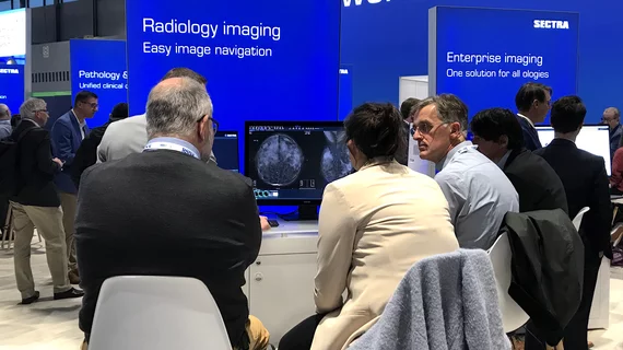
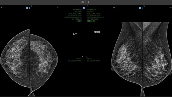
Picture archiving and communication systems (PACS) have replaced conventional radiographic films as the digital image-viewing hub over the past two decades and now serve as the primary communication bridge between radiologists, radiologic technologists and referring providers. PACS enables all authorized clinicians to access medical images and reports quickly, easily and from virtually any location. Some health systems have integrated PACS into the electronic medical record (EMR). Others have moved to enterprise image systems to replace radiology PACS and allow all departments to now store images and reports in one location for easier health system-wide access.

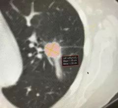
Example of artificial intelligence generated measurements to quantify the size of a lung cancer nodule during a followup CT scan to see if the lesion is regressing with treatment. This type of automation can aid radiologists by doing the tedious, time consuming work. Photo by Dave Fornell

8 trends in radiology technology to watch in 2023

Here is a list of some key trends in radiology technology from our editors based on our coverage of the radiology market.

January 18, 2023
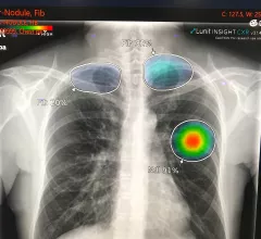
Example of AI automated detection and highlighting of critical lung findings on a chest X-ray for a possible lung cancer nodule and fibrosis. Example shown by AI vendor Lunit.

VIDEO: Radiology AI trends at RSNA 2022

Sanjay Parekh, PhD, senior market analyst with Signify Research, discusses trends in radiology AI seen on the expo floor and in sessions at RSNA 2022.

December 12, 2022
RIS/PACS: Driving Standardization for a Large Hospital System

PocketHealth launches network-free image sharing and storage platform

PocketHealth has launched a platform that lets patients and providers securely request, share and store medical images without the use of CD-ROMS or networks.

November 14, 2022

University of Rochester Medical Center in the US selects Sectra Enterprise Imaging in the cloud

Linköping, Sweden and Shelton, CT – October 17, 2022– International medical imaging IT and cybersecurity company Sectra (STO: SECT B) will provide enterprise imaging as a cloud subscription service (Sectra One Cloud), throughout the University of Rochester Medical Center (URMC). This will allow the US health system scalability as enterprise imaging volumes grow, in a secure and fully managed cloud environment.

October 19, 2022
Monique Rasband, vice president of imaging, cardiology and oncology, KLAS Research, explains some of technology trends KLAS researchers have found in enterprise imaging system and radiology artificial intelligence (AI).

VIDEO: KLAS shares trends in enterprise imaging and AI

Monique Rasband, vice president of imaging, cardiology and oncology, KLAS Research, explains some of technology trends KLAS researchers have found in enterprise imaging system and radiology artificial intelligence (AI).

October 13, 2022
Charles E. Kahn, Jr., MD, MS, Editor of the the journal Radiology: Artificial Intelligence, and professor and vice chair of radiology at the University of Pennsylvania Perelman School of Medicine. He has been heavily involved in radiology informatics and has seen up close the evolution of radiology toward deeper integration with AI. #RSNA

VIDEO: Use cases and implementation strategies for radiology artificial intelligence

Charles Kahn, Jr., MD, editor of the the journal Radiology: Artificial Intelligence, and professor and vice chair of radiology at the University of Pennsylvania Perelman School of Medicine, explains the work involved integrating AI in radiology systems and the role of AI in augmenting patient care.
 

October 12, 2022
Monique Rasband from KLAS Research shares trends in PACS and radiology informatics.

VIDEO: 6 key trends in PACS and radiology informatics observed by KLAS

Monique Rasband, vice president of imaging, cardiology and oncology, KLAS Research, shares some of technology trends observed in radiology PACS and and imaging informatics since 2019.

September 29, 2022
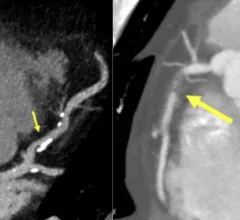
Examples of new plaque reporting in the CAD-RADS 2.0 document. Left, an example from CAD-RADS 2 / P2 plaque burden with mild coronary stenosis (25-49%). Right, example of a CAD-RADS 5/ P3, with a focal, non-calcified occlusion of the proximal RCA (arrow) and severe amount of plaque (P3). #CADRADS #YesCCT #CTA #CCTA

New CAD-RADS 2.0 reporting for coronary CTA offers patient management recommendations

The document includes updated classification to established a framework for stenosis, plaque burden and plaque modifiers, including assessment of CT-FFR or myocardial CT perfusion.

July 8, 2022

Around the web

Automated AI-generated measurements combined with annotated CT images can improve treatment planning and help referring physicians and patients better understand their disease, explained Sarah Jane Rinehart, MD, director of cardiac imaging with Charleston Area Medical Center.

Two advanced algorithms—one for CAC scores and another for segmenting cardiac chamber volumes—outperformed radiologists when assessing low-dose chest CT scans. 

"Gen AI can help tackle repetitive tasks and provide insights into massive datasets, saving valuable time," Thomas Kurian, CEO of Google Cloud, said Tuesday. 

Trimed Popup
Trimed Popup