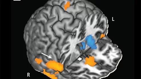
Cocaine addiction can ruin a person physically and financially, and with an estimated 1.4 million cocaine users in the U.S., thousands will become trapped by their habit. While previous research on the drug and its addictive potential were observational and subjective, imaging is reshaping how we see addiction—and how it will be treated.