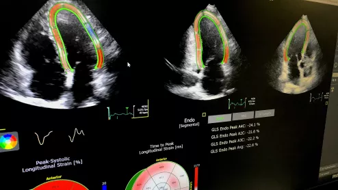
Despite its diagnostic and prognostic value, speckle-tracking strain echocardiography is underused, some cardiac imagers say. What will it take for adoption to pick up?
With the “volume to value” movement pushing radiologists to prove their contributions to cost containment, some are feeling uneasy. After all, imaging utilization stands to be curbed—or at least eyed more closely than ever before for appropriateness.
What should radiology be expending, in manpower as well as money, to help make medical imaging accessible to and from every clinical department? And what’s in enterprise imaging for radiology, anyway?
Cardiologists are receiving more exposure to different imaging modalities during their fellowships, but their job prospects and training vary widely. A more comprehensive and multimodality training approach could lead to better results.
Physicians in fields like cardiology have traditionally looked to clinical practice guidelines to help articulate the best evidence-based care for patients. The rapidly growing movement to value-based care is prompting clinicians—including echocardiographers—to carefully weigh a more focused and integrative approach to delivering consistent, quality medicine: care pathways.
Treating today’s cancer patient no longer means simply targeting the cancer. Given the known cardiotoxicities of some established chemotherapies and the possibility that newer approaches may damage the heart, oncologists, cardiologists and imaging specialists now work together to detect and minimize the risk of treatment-induced heart failure.
Cardiovascular information systems (CVIS) are providing the means to integrate the business of cardiology with the practice of cardiology at the point of care—with great results for managers.
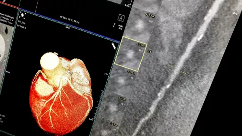
Automated AI-generated measurements combined with annotated CT images can improve treatment planning and help referring physicians and patients better understand their disease, explained Sarah Jane Rinehart, MD, director of cardiac imaging with Charleston Area Medical Center.
Two advanced algorithms—one for CAC scores and another for segmenting cardiac chamber volumes—outperformed radiologists when assessing low-dose chest CT scans.