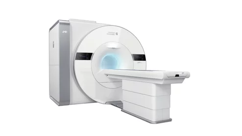
Houston, Texas-based United Imaging announced the clearance of its uMR Jupiter 5T MRI system, the first to offer an 8-channel whole-body multi-transmit system.
The model was developed and validated specifically for liver, spleen and pancreas segmentation, and outperformed a publicly available segmentation model already in use.
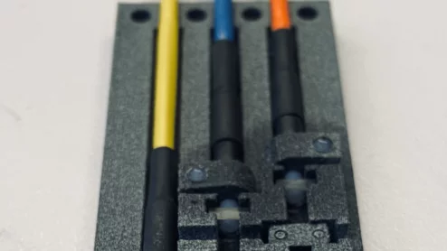
The sensor uses laser light encased in fiber cables and a small glass container filled with gas to measure changes in the strength of a magnetic field.
This latest research further confirms that breast MRI not only detects tumors that mammography cannot, but it also spots invasive cases that pose greater risks to patients.
Although these tools have proven themselves valuable in numerous settings, they must be used with caution, especially by patients and nonradiologist providers who may be seeking clarification on imaging reports.
A general lack of awareness pertaining to ACR appropriateness criteria could be a driving factor behind the misguided requisitions, authors of a new analysis suggest.
Experts, medical organizations and advocates alike are coming forward saying that the new guidelines “do not go far enough,” particularly when it comes to addressing the screening needs of certain patients.
Society of Cardiovascular Computed Tomography (SCCT) President Brian Ghoshhajra, MD, detailed three challenges he is making to SCCT members in the coming year.
The authors stated that their results should not deter providers from offering their patients radiotherapy treatment but rather encourage in-depth discussions and shared decisions pertaining to the best options.
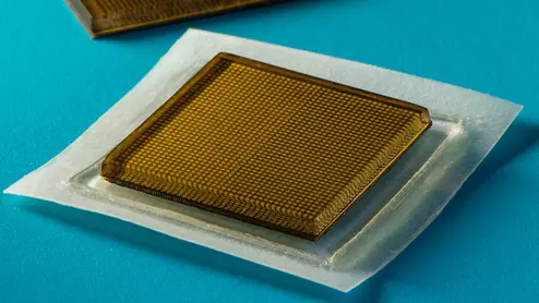
Experts at MIT have developed ultrasound stickers that can be worn in the same manner as a Band-Aid while also producing diagnostic quality images in real-time.
Automated AI-generated measurements combined with annotated CT images can improve treatment planning and help referring physicians and patients better understand their disease, explained Sarah Jane Rinehart, MD, director of cardiac imaging with Charleston Area Medical Center.
Two advanced algorithms—one for CAC scores and another for segmenting cardiac chamber volumes—outperformed radiologists when assessing low-dose chest CT scans.