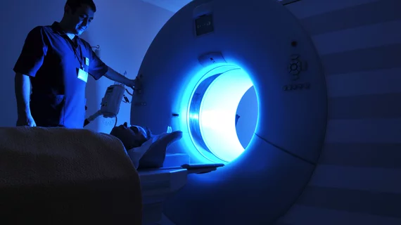
Researchers from Brigham Young University in Provo, Utah were able to complete MRI scans of more than 30 children and adolescents with autism and low verbal and cognitive performance (LVCP) without the use of sedation by using behavioral support procedures to reduce patient fear and anxiety.
The team, led by Terisa P. Gabrielsen, PhD, assistant professor of counseling psychology and special education at Brigham Young, successfully conducted structural and functional MRI (fMRI) scans of 37 children and adolescents between the ages of 7 and 17 years with autism—including 17 with less-developed language skills and an average IQ of 54, according to research published online Jan. 2 in the journal Molecular Autism.
“This is such an overlooked group of kids and adults,” co-author Ryan Kellems, PhD, assistant professor of Counseling Psychology and Special Education, said in a prepared statement. “We don’t know very much about their brains because people say they are challenging to study.”
Prior to their scans, the researchers gave the children and their families a video to watch at home explaining step-by-step the fMRI process and audio files to prepare them for the sounds they’d hear in the machine.
At the time of their scan, the children could push buttons on the MRI machine to move it up and down. During the scan, the researchers reassured the children by holding their hand, reminding them to relax and allowing them to wear noise-cancelling headphones.
After the scans were complete, the researchers analyzed how the children from the LVCP group compared to higher performance children with autism as well as neurotypical children.
Within the LVCP group, some children’s brain’s networks had decreased activity between the left and right hemispheres, according to the researchers. Network connectivity, however, was higher for the LVCP group than for the neurotypical group.
Although a child with autism and LVCP may not possess a wide range of language skills, it’s never safe to assume that their brain in underactive, the researchers noted. Rather, their brain may be more active depending on what it’s attending to.
“Our findings point to the influence of non-diagnostic factors, such as cognitive ability for understanding brain connectivity in autism and other neurodevelopmental disorders, while highlighting some similarities that may be consistent throughout autism regardless of level of verbal and cognitive ability,” the researchers wrote. “This project emphasizes the need for studying the entire spectrum of autism in order to elucidate individual difference factors related to etiology and outcome and to begin to develop more specific targets for intervention towards improved outcomes.”

