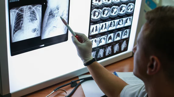
High-resolution computed tomography imaging revealed age-related differences in lung abnormalities among patients with the coronavirus. Experts believe their insights can help develop tailored treatment strategies to combat the growing pandemic.
A crew of Chinese scientists analyzed case data and chest imaging taken from nearly 100 patients, ages 4 to 88 years old, with COVID-19. They identified ground-glass opacities as the most common finding, with individuals 45 and older showing more lesions than those younger than 18 years old.
Importantly, nearly 80% of the patients in this study had a history of direct or indirect contact with people from Wuhan, China—the center of the outbreak—the authors noted March 26 in the European Journal of Radiology. But the team still believes their research can help other institutions, including radiology departments.
“In this study, the key finding is that the middle-aged and elderly patients have the most severe representation of parenchymal findings, and follow with the morphological appearance that seems to be particularly limited in youngest group, which provides certain support for clinical individualized treatment of patients of different ages,” wrote Zuhua Chen, with Hangzhou Xixi Hospital’s Department of Radiology, and colleagues.
Although CT imaging is not currently recommended as a first-line screening tool in suspected COVID-19 cases, other research has reported that the modality can be useful for stratifying patients according to disease severity and assist in personalizing treatment.
For their study, Chen et al. had two expert radiologists look over 98 high-resolution CT scans taken from the hospital’s picture archiving and communication system. They separated the group by age into four different cohorts.
Overall, the most common abnormalities were small, patchy ground-glass opacities and consolidations. Individuals ages 45-59 and those older than 60 years, however, demonstrated more involvement in both lungs, the lung lobe and lung field; these patients also had a higher lesion count compared to people under the age of 18.
“Our study findings showed that middle-aged and older patients had more severe lung involvement and lobe involvement and, at the same time, the lesions were accompanied more often by air bronchograms,” the authors noted.
Chen and colleagues suggested a few possible causes for their discoveries, including the weak “virulence” of COVID-19 that results in lighter imaging signs in younger patients, and the likelihood that older patients have more baseline diseases making them more vulnerable to an attack on the immune system.
The study was limited by its small number of adolescent patients and the fact that imaging merely depicts a single second in a patient’s development of the new virus; follow-up imaging could yield different abnormalities.
“These study findings are likely to help with understanding and evaluating the condition of infected patients of different ages, and provide insight into the possible subsequent evolution of the disease in infected patients of different ages, which should facilitate more accurate diagnosis and the development of treatment strategies,” the authors concluded.

Matt joined Chicago’s TriMed team in 2018 covering all areas of health imaging after two years reporting on the hospital field. He holds a bachelor’s in English from UIC, and enjoys a good cup of coffee and an interesting documentary.