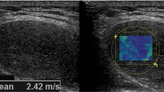
Shear-wave elastography (SWE) may help distinguish musculoskeletal soft-tissue lesions as benign or malignant in conjunction with conventional ultrasound, according to research published online Nov. 27 in Radiology.
An emerging imaging technique that uses ultrasound to identify the biomechanical properties of tissue, SWE measures the velocity of propagation of shear waves produced by the transducer to determine the elasticity of tissues.
However, SWE did not improve the diagnostic performance for lesions when MRI was combined with ultrasound. Additionally, the association between shear-wave velocity (SWV) and malignancy varied by lesion location and the patient’s age, according to Philip Robinson, MD, radiologist at Leeds Teaching Hospitals NHS Trust in the U.K., and colleagues.
“Although SWE does not have independent predictive ability, its supplemental use is able to improve the classification performance of assessment using ultrasound alone when compared with histologic evaluation,” Robinson et al. wrote.
Recruited for the study from December 2015 through March 2017 were 206 participants (average age 58 years, 57 percent women) who were scheduled for biopsy of a soft-tissue mass. All underwent ultrasound, MRI and SWE prior to their biopsies.
Ultrasound images alone, and ultrasound and MRI images together were independently reviewed by three musculoskeletal radiologists who classified lesions as benign, probably benign, probably malignant or malignant.
For SWE, the area under the receiver operating characteristic (ROC) curve (AUC) was calculated for transverse SWV and multivariable logistic regression was used to investigate the association between SWE and malignancy alongside individual demographic and imaging variables, according to the researchers.
Of the total number of participants, 38 percent had malignant lesions. The researchers found that SWV with ultrasound had an AUC of 0.87 and a 95 percent confidence interval, showing an overall good diagnostic accuracy for classifying lesions as benign or probably benign.
However, SWE did not perform with comparable diagnostic accuracy with or without MRI in classifying lesions as probably malignant or malignant.
“Given the ubiquity of lipomatous lesions and the difficulties in managing them, further evaluation of the role of shear-wave elastography in lesion classification should be performed, especially given that its most powerful ability appears to be adding credence to the impression of benignity based on ultrasound alone,” the researchers concluded.

