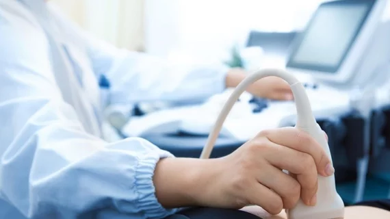
At many institutions, ultrasound is preferred for imaging patients with suspected appendicitis, but highly trained operators are typically unavailable during off hours. A new study, however, suggests radiology residents may be able to fill this gap in care.
Physicians from the University of British Columbia in Vancouver, Canada, compared more than 200 scans performed by sonographers and residents at their hospitals, sharing their results Aug. 14 in Academic Radiology.
Both groups performed similarly, but on-call residents completed a greater number of focused exams (missing key organs) which led to an uptick in downstream CT and MRI, the authors noted.
“Based on our findings it is suggested that US scans performed for suspected appendicitis in this patient population attempt to assess the right kidney, right ovary and terminal ileum as we discovered an increased rate of downstream imaging in cases in which residents performed limited scans and did not document visualization of the pelvic organs,” James M. Roberts, MD, MSc, with the university’s radiology department, and colleagues wrote Friday.
Acute appendicitis is the most common non-obstetric surgical emergency, the authors noted, with 104 cases per 100,000 person-years in women between 13 and 40 years old. And the ACR Appropriateness Criteria suggests using ultrasound in these situations.
Roberts et al. set out to test the scanning practices of on-call residents who perform urgent US outside of normal working hours, against trained sonographers operating during the day.
After comparing 104 scans from residents to the same number completed by sonographers, the latter group visualized the appendix in slightly more cases (30% versus 27%). Both parties’ sensitivity and specificity for appendicitis also proved similar: Each notched a 98% specificity, while sonographers edged out on-call residents in sensitivity, 69% vs. 63%, respectively.
Residents did not image pelvic organs in 34% of cases, far surpassing sonographer’s mark of 1%. And in these focused scanning situations, follow-up imaging utilization was more likely.
The authors noted there isn’t a standardized approach for reporting cases in which the appendix is not visible, and suggested downstream imaging could be impacted by the reporting radiologist, which should be studied.
“Out-of-hours US scans performed by radiology residents had similar performance characteristics compared to departmental sonographers,” the authors concluded. “We discovered an increased rate of downstream imaging in cases in which residents performed limited scans and did not document visualization of the pelvic organs.”

Matt joined Chicago’s TriMed team in 2018 covering all areas of health imaging after two years reporting on the hospital field. He holds a bachelor’s in English from UIC, and enjoys a good cup of coffee and an interesting documentary.