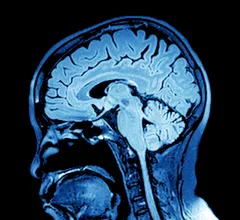
Magnetic Resonance Imaging

Magnetic resonance imaging (MRI) is the gold standard imaging modality for soft tissues. It produces detail cross-sectional images of soft tissue and bone anatomy, including muscles, tendons, ligaments, brain and organs, without the use of ionizing radiation. In addition to orthopedic imaging, MRI is also used for heart, brain and breast. MRI uses gadolinium contrast in many exams to highlight tissues and blood vessels, which enhances images and offers better diagnostic quality. It can also be used in conjunction with PET scans. How does MRI work? MR creates images by using powerful magnets to polarize hydrogen atoms in water (the body is made of of more than 80% water) so they face in one direction. A radiofrequency pulse is then used to ping these atoms, causing them to wobble, or resonate. The MRI coils detect this and computers can assemble images from the signals. Basic MRI scans will focus on the resonance of fat and water in two different sequences, which highlight and contrast different features in the anatomy.
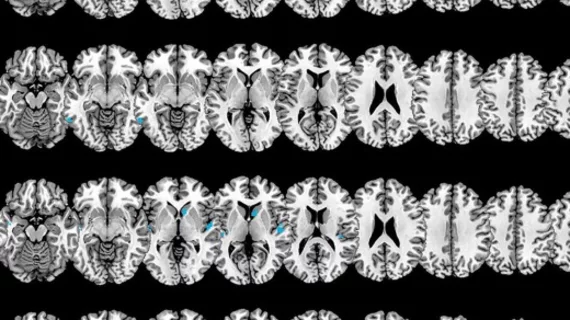
MRI-based measurements can predict urinary continence after prostate surgery

Radiologists should complete specific training to understand these measurements before using membranous urethra length as a predictor of postoperative continence, University of Michigan researchers suggested.

January 19, 2022

6 tips for making MRI exams more autism-friendly

Clinicians should focus on ensuring exams are more compatible with autism patients rather than making such individuals more MR-compatible.

January 17, 2022

Abbreviated MRI outperforms DBT for detecting secondary breast cancer

“Our study results suggest that AB-MRI could potentially be sufficient for post-treatment surveillance,” experts suggested Thursday in Academic Radiology.

January 13, 2022

Researchers are developing a modified MRI protocol to help treat brain hemorrhages

“We are studying whether MRI can provide more complete information which could alter the clinical management of patients with hemorrhagic stroke," an expert involved in the trial explained.

January 12, 2022

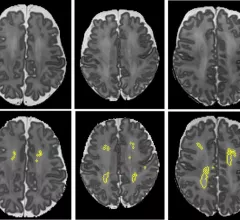
Doctors suggest performing whole-spine MRI scans for suspected child abuse victims

Replacing cervical spine exams with whole-spine MRIs can reveal "major findings," investigators explained recently.

January 12, 2022
Robot Eye

Combining functional MRI scans with eye tests could help stroke survivors recover lost vision

Identifying the exact location of the damage in a patient's visual pathway may enable doctors to develop unique rehabilitation methods,  experts explained in Frontiers in Neuroscience.

January 7, 2022

Doctors pilot mobile stroke unit equipped with low-cost MRI machine

Medical University of South Carolina experts believe they are the first to place a portable magnetic resonance imaging unit inside of an ambulance.

January 6, 2022
Brain MRI

New MRI technique earlier detects multiple sclerosis, potentially improving treatment approaches

The tool identified amino acid derivatives that are linked to compromised neuron integrity.

January 4, 2022

Around the web

Automated AI-generated measurements combined with annotated CT images can improve treatment planning and help referring physicians and patients better understand their disease, explained Sarah Jane Rinehart, MD, director of cardiac imaging with Charleston Area Medical Center.

Two advanced algorithms—one for CAC scores and another for segmenting cardiac chamber volumes—outperformed radiologists when assessing low-dose chest CT scans. 

"Gen AI can help tackle repetitive tasks and provide insights into massive datasets, saving valuable time," Thomas Kurian, CEO of Google Cloud, said Tuesday. 

Trimed Popup
Trimed Popup