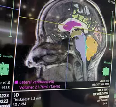
Magnetic Resonance Imaging

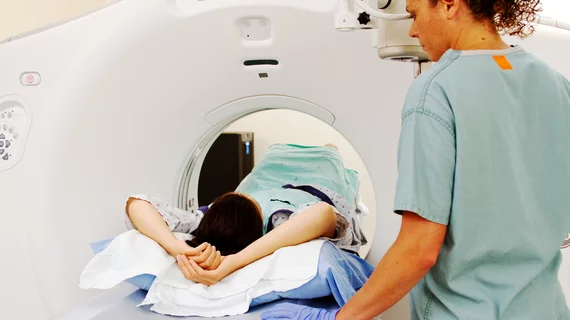
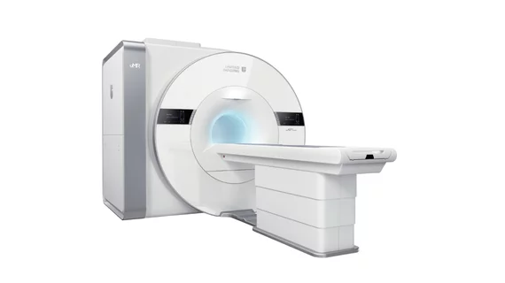
Magnetic resonance imaging (MRI) is the gold standard imaging modality for soft tissues. It produces detail cross-sectional images of soft tissue and bone anatomy, including muscles, tendons, ligaments, brain and organs, without the use of ionizing radiation. In addition to orthopedic imaging, MRI is also used for heart, brain and breast. MRI uses gadolinium contrast in many exams to highlight tissues and blood vessels, which enhances images and offers better diagnostic quality. It can also be used in conjunction with PET scans. How does MRI work? MR creates images by using powerful magnets to polarize hydrogen atoms in water (the body is made of of more than 80% water) so they face in one direction. A radiofrequency pulse is then used to ping these atoms, causing them to wobble, or resonate. The MRI coils detect this and computers can assemble images from the signals. Basic MRI scans will focus on the resonance of fat and water in two different sequences, which highlight and contrast different features in the anatomy.

Kid holding baseball

New radiology research may shed light on MLB’s record number of Tommy John surgeries

A retrospective analysis of elbow MRI exams shows young baseball players can develop lasting injuries.

December 13, 2023
Damaged Organ

AI model with single-source dataset outperforms multi-institution version

Despite the results, researchers still recommend using models trained on multi-institutional datasets.

December 12, 2023
Hacker

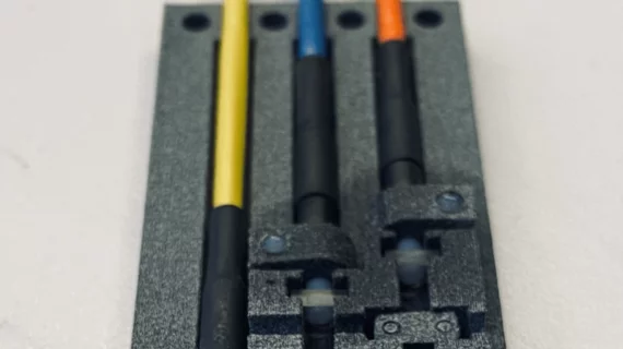
'Hackers' created a fully functional MRI machine in four days

The proof of concept is available online as 3D-printable files.

December 11, 2023
Brain MRI AI assessment and segmentation on Fujifilm's Synapse system at RSNA 2023. Photo by Dave Fornell. #RSNA #RSNA23 #RSNA2023

PHOTO GALLERY: New technology at RSNA 2023

Images from the world's largest radiology conference include new technologies and the latest advances in MRI, CT, nuclear medicine, X-ray, artificial intelligence, and PACS/enterprise imaging.

December 11, 2023
handgun.jpg

Woman shot in buttocks by own gun during MRI appointment

The unnamed woman had concealed the weapon, which the MRI machine triggered to fire during an exam. 

December 4, 2023
offender-trouble-handcuffs-police-arrest-2102488.jpg

Imaging chain faces regulators after inmate, guard get stuck to MRI machine

After an investigation by journalists, unregulated SimonMed locations in Arizona must now obtain a license.

December 1, 2023
Philips Vanderbilt Carbon Emissions Warning

How radiology departments can decarbonize, save money

Philips and Vanderbilt University Medical Center have released findings from their efforts to reduce waste in the hospital's radiology department.

December 1, 2023
CAIRE-MRI Portal screenshot

Company launches world's first online portal for MRI safety reporting

The web-based portal aims to log all accidents and any associated remedies, bringing as much information as possible to a central location.

November 16, 2023

Around the web

Automated AI-generated measurements combined with annotated CT images can improve treatment planning and help referring physicians and patients better understand their disease, explained Sarah Jane Rinehart, MD, director of cardiac imaging with Charleston Area Medical Center.

Two advanced algorithms—one for CAC scores and another for segmenting cardiac chamber volumes—outperformed radiologists when assessing low-dose chest CT scans. 

"Gen AI can help tackle repetitive tasks and provide insights into massive datasets, saving valuable time," Thomas Kurian, CEO of Google Cloud, said Tuesday. 

Trimed Popup
Trimed Popup