Nuclear Medicine

Nuclear medicine (also called molecular imaging) includes positron emission computed tomography (PET) and single photon emission computed tomography (SPECT) imaging. Nuclear imaging is achieved by injecting small amounts of radioactive material (radiopharmaceuticals) into patients before or during their scan. These can use sugars or chemical traits to bond to specific cells. The radioactive material is taken up by cells that consume the sugars. The radiation emitted from inside the body is detected by photon detectors outside the body. Computers take the data to assemble images of the radiation emissions. Nuclear images may appear fuzzy or ghostly rather than the sharper resolution from MRI and CT.  But, it provides metabolic information at a cellular level, showing if there are defects in the function of the heart, areas of very high metabolic activity associated with cancer cells, or areas of inflammation, data not available from other modalities. These noninvasive imaging exams are used to diagnose cancer, heart disease, Alzheimer’s and Parkinson’s disease, bone disorders and other disorders. 

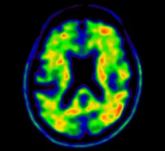
An example of a positive amyloid plaque PET nuclear brain scan. these tests can help identify Alzheimer's patients earlier so they can be prescribed drugs to treat the condition.

A new Alzheimer's drug therapy appears effective. What might this mean for the future of amyloid PET?

Previously, CMS determined that coverage for patients receiving treatment was dependent on their being enrolled in a CMS-approved clinical trial under coverage with evidence development (CED).

September 29, 2022
A study published this week in the Journal of the American College of Cardiology (JACC): Cardiovascular Imaging shows artificial intelligence (AI) algorithms can more rapidly and objectively determine calcium scores in computed tomographic (CT) and positron emission tomographic (PET) images than physicians.[1] The AI also performed well when the images were obtained from very-low-radiation CT attenuation scans. https://doi.org/10.1016/j.jcmg.2022.06.006

Artificial intelligence can objectively determine cardiac calcium scores faster than doctors

A new study shows artificial intelligence (AI) algorithms can more rapidly and objectively determine calcium scores in CT and PET/CT images than physicians.

September 16, 2022
Prostate Cancer

Does the use of PSMA PET impact tumor upstaging?

A new risk analysis suggests the exam will increasingly result in tumor upstaging among high-risk patients.

September 15, 2022
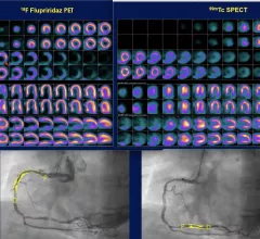
Comparison of flurpiridaz F-18 PET, SPECT and angiography of the 60-year-old female patients in the Aurora trial. The SPECT scan appeared normal, but flurpiridaz was shown to be more sensitive and showed the ischemia from two blockages in the right coronary artery. The new radiotracer may help expand cardiac PET. #ASNC

Flurpiridaz data shows promise to expand and enhance cardiac PET

The biggest news from the American Society of Nuclear Cardiology (ASNC) 2022 meeting was positive late-breaking data on the phase 3 Aurora trial for the flurpiridaz (F-18) PET radiotracer agent.

September 12, 2022

FDG-PET shown to predict pancreatic cancer outcomes prior to surgery, could guide treatment decisions

FDG-PET scans in these patients allow clinicians to determine whether the tumors are still viable or not, thus playing a significant role in making treatment decisions.

September 9, 2022
Two of the top stories this month included the death of MRI pioneer Raymond Vahan Damadian. Experts at MIT also developed ultrasound stickers that can be worn in the same manner as a Band-Aid while also producing diagnostic quality images in real-time. #MRI

Top Health Imaging stories in August 2022

Here are the most popular stories on Health Imaging in August, based on our website analytics. 

September 1, 2022
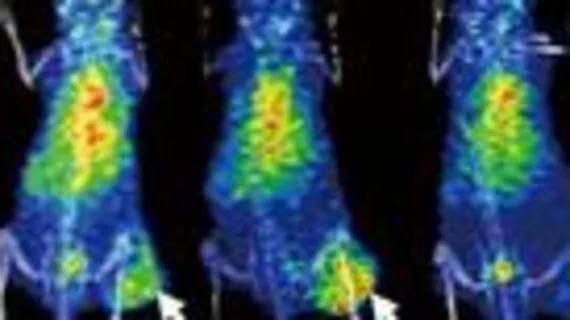
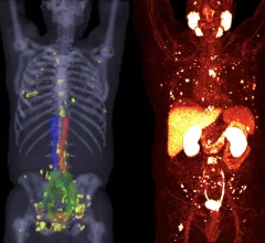
Two examples of PSMA-PET scans showing numerous prostate cancer metastases spread throughout the body. Many of these smaller tumors would not have been detected on previous standard-of-care imaging. Photo on left courtesy of SNMMI, right University of Chicago. #PSMAPET

PSMA-PET is rapidly changing the standard of care for prostate cancer patients

Adoption of PSMA-PET has been swift because it can significantly improve prostate cancer detection and treatment. SNMMI President Munir Ghesani, MD, explains how.

August 26, 2022
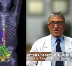
Munir Ghesani, MD, SNMMI president, explained the impact of PSMA-PET imaging on nuclear image and treatment for prostate cancer.

VIDEO: Advantages of PSMA-PET imaging in prostate cancer care

Munir Ghesani, MD, president of the Society of Nuclear Medicine and Molecular Imaging (SNMMI) and system chief of nuclear medicine at Mount Sinai Health, explains PSMA-PET has seen rapid adoption because it can significantly improve prostate cancer detection and treatment. 
 

August 25, 2022

Around the web

Automated AI-generated measurements combined with annotated CT images can improve treatment planning and help referring physicians and patients better understand their disease, explained Sarah Jane Rinehart, MD, director of cardiac imaging with Charleston Area Medical Center.

Two advanced algorithms—one for CAC scores and another for segmenting cardiac chamber volumes—outperformed radiologists when assessing low-dose chest CT scans. 

"Gen AI can help tackle repetitive tasks and provide insights into massive datasets, saving valuable time," Thomas Kurian, CEO of Google Cloud, said Tuesday. 

Trimed Popup
Trimed Popup