Echocardiography

Cardiac ultrasound uses reflected sound waves (echos) to create images of anatomy inside the body. Echocardiograms are the primary cardiac imaging modality used to assess the heart and diagnose or track cardiac issues. Echo is the gold standard imaging modality to assess the heart, particularly with calculating left ventricular ejection fraction (LVEF), which is a measure of cardiac output. In addition to noninvasive standard transthoracic echo (TTE), invasive transesophgeal echo (TEE) is also used when clearer, more detailed imaging of the heart is needed. Both 3D and 4D echo echo systems are rapidly gaining wider adoption and enable new types of assessments, especially in the structural heart space and in transcatheter procedural guidance. Find news on general ultrasound imaging.

artificial intelligence consultation

Imaging specialists partner with Pfizer to deliver AI-powered cardiac amyloidosis evaluations

Improving care for patients with cardiac amyloidosis has emerged as one of the hottest topics in cardiology. Pfizer helped kickstart that trend in 2019 when it gained FDA approval for two separate medications for the rare, but potentially fatal disease. 

January 10, 2024
transesophageal echocardiography (TEE)

New expert guidelines: Start PHV evaluations with echocardiography, but other imaging modalities can provide value

New guidelines from the American Society of Echocardiography, made in collaboration with the Society for Cardiovascular Magnetic Resonance and Society of Cardiovascular Computed Tomography, represent an update of the group's original recommendations from 2009.

January 9, 2024
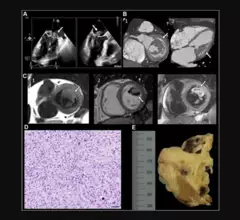
“Undifferentiated Cardiac Sarcoma of the Mitral Valve: Multimodal Imaging Assessment" Radiology: Cardiothoracic Imaging RSNA

Radiologists select 2023's top images in cardiothoracic imaging

“Cardiothoracic imaging continues to evolve, fueled by technical innovations such as dynamic chest radiography, digital tomosynthesis and dark-field radiography," one radiologist said.

December 21, 2023
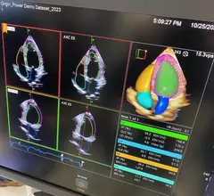
Example of a totally AI driven echocardiography workflow on the new Siemens Origin ultrasound system unveiled in 2023. The AI did all the work on this screen, taking a 3D echo exam and automatically segmenting the anatomy, contoured all the chambers, found the ideal views to display and then calculated all the measurements in seconds. Photo by Dave Fornell at TCT 2023. 

Cardiology now has more than 100 FDA cleared AI algorithms

Cardiology makes up 10% of the 692 market-cleared clinical AI algorithms in the FDA’s latest update on the number of patient-facing AI now commercialized in the U.S. Experts share their thoughts on how it is being used.

December 14, 2023
artificial intelligence AI heart cardiology

ChatGPT struggles with echocardiography, but still shows potential to help cardiology trainees

ChatGPT may not be quite ready to help prepare trainees for their next echocardiography exam—but it does show promise. 

November 16, 2023
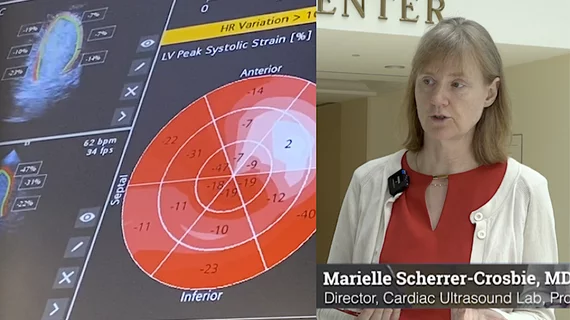
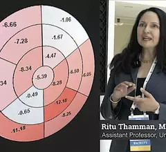
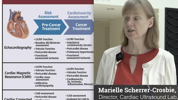
Ritu Thamman, MD, FASE, University of Pittsburgh School of Medicine, discusses trends and the technical aspects of strain imaging. #ASE #ASE23 #ASE2023 #strainecho

Strain imaging making a significant impact on the treatment of heart patients

Strain imaging has gained considerable momentum in recent years as an effective tool for getting more value out of heart evaluations. Ritu Thamman, MD, provided an in-depth look at the latest trends in strain echocardiography in an exclusive interview. 

September 14, 2023
Video of Denise Garris, ASE regulatory consultant and principal of the Korris Group, explaining how reimbursements from insurance andMedicare work to pay or cardiac ultrasound exams. #ASE #ASE360 #ASE23 #ASE2023 #healthcarereimbursment

Understanding reimbursements and coding for echocardiography

"I think everyone would agree that the current fee-for-service model is not working appropriately and there needs to be a fix," regulatory consultant Denise Garris explained in a new interview. 

September 13, 2023
Arzhang Fallahi, MD, and David Hsi, MD, discussing imaging-based aortic stenosis screening

Q&A: Cardiologists explore the potential impact of a screening program for aortic stenosis

We already screen patients for breast cancer and lung cancer on a regular basis. Why not establish screening programs for aortic stenosis?

September 11, 2023

Around the web

Automated AI-generated measurements combined with annotated CT images can improve treatment planning and help referring physicians and patients better understand their disease, explained Sarah Jane Rinehart, MD, director of cardiac imaging with Charleston Area Medical Center.

Two advanced algorithms—one for CAC scores and another for segmenting cardiac chamber volumes—outperformed radiologists when assessing low-dose chest CT scans. 

"Gen AI can help tackle repetitive tasks and provide insights into massive datasets, saving valuable time," Thomas Kurian, CEO of Google Cloud, said Tuesday. 

Trimed Popup
Trimed Popup