Cardiac Imaging

While cardiac ultrasound is the widely used imaging modality for heart assessments, computed tomography (CT), magnetic resonance imaging (MRI) and nuclear imaging are also used and are often complimentary, each offering specific details about the heart other modalities cannot. For this reason the clinical question being asked often determines the imaging test that will be used.
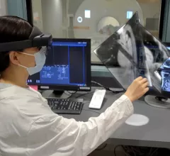
mixed reality LAAO Chase Western Reserve MRI. The group—which includes engineers, cardiologist, radiologists and other specialists—will attempt to perform a robotic-controlled left atrial appendage occlusion (LAAO) on a patient inside an MRI scanner.

Researchers receive $3.7M to attempt robotic heart surgery inside MRI scanner

The group—which includes engineers, cardiologist, radiologists and other specialists—will attempt to perform a robotic-controlled left atrial appendage occlusion (LAAO) on a patient inside an MRI scanner.

October 20, 2022
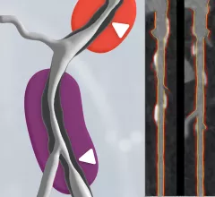
Left, HeartFlow's RoadMap analysis enables cardiac CT readers to identify stenoses in the major coronary arteries. The AI provides visualization and quantification of the location and severity of anatomic narrowings. Right image, HeartFlow's Plaque Analysis AI algorithm automates assessment of coronary plaque characteristics and volume on CCTA exams to greatly reduce the time it takes to manually assess and quantify these features.

HeartFlow gains FDA clearance for 2 new AI-powered imaging assessments

The solutions, Plaque Analysis and RoadMap Analysis, both use coronary CT angiography to provide clinicians with a noninvasive look at patients who present with coronary artery disease and face a heightened myocardial infarction risk.

October 18, 2022
Coronary CT Angiography (CCTA) has evolved into a much more helpful resource than interventional cardiologists may have originally believed. In fact, the Society of Cardiovascular Computed Tomography (SCCT) emphasized in a new expert consensus statement that CCTA provides specialists with an effective tool in the preparation and optimization of coronary procedures. The 2021 chest pain guidelines elevated CTA to a class 1A recommendation. Harold Litt , MD, explains the use of CCTA in the ED.

VIDEO: Cardiac CT as a front-line chest pain exam in the ED

Harold Litt, MD, explains the use of cardiac CT as a primary imaging exam for chest pain at his emergency department over the past decade.

October 13, 2022
Chest pain

FAPI PET/CT findings linked with risk of sudden cardiac death

Experts involved in the research suggested that these findings indicate a potential role for FAPI PET/CT imaging in detecting changes in myocardial fibrosis, noting that it could be more sensitive to earlier changes than standard cardiac MRI.

October 12, 2022
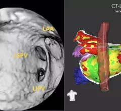
CT has been used for planning and procedural guidance in pulmonary vein isolation (PVI) for about 20 years. It shows the anatomy to the electrophysiologist. The EPs also can load the imaging into their EP mapping system. It also shows them the location of the esophagus and the phrenic nerve so they can avoid these during the procedures. #PVI #EPeeps

VIDEO: The expanding role of cardiac CT in electrophysiology

Harold Litt, MD, division chief of cardiothoracic imaging and director of the Center for Advanced CT Imaging Sciences at the Perelman School of Medicine of the University of Pennsylvania, explains the role of cardiac CT in EP procedures.

October 12, 2022
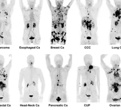
FAPI PET/CT shows promise as a better radiotracer for cancer than 18F-FDG. It also can be used for detection of infection and inflammation.

VIDEO: 6 key advances in molecular imaging technology for PET and SPECT

Munir Ghesani, MD, President of the Society of Nuclear Medicine and Molecular Imaging (SNMMI), system chief of nuclear medicine at Mount Sinai Health, explains recent advances in nuclear imaging technology.

September 30, 2022

New recommendations highlight the importance of CCTA when treating acute chest pain in the ED

The Society of Cardiovascular Computed Tomography developed and published the new document to help educate healthcare providers who regularly treat acute chest pain in the emergency department. 

September 29, 2022
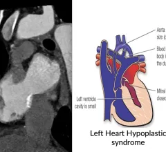
CT imaging showing the congenital heart defect of left heart hypoplastic syndrome.

VIDEO: Congenital heart imaging with cardiac CT

As the population of patients with adult congenital heart disease grows, they are presenting to adult cardiology clinics and being imaged with CT. Many also do not have access or cannot be imaged by MRI, said Renee Bullock-Palmer, MD.

September 28, 2022

Around the web

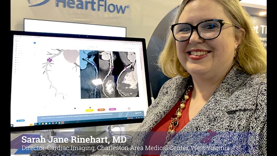
Automated AI-generated measurements combined with annotated CT images can improve treatment planning and help referring physicians and patients better understand their disease, explained Sarah Jane Rinehart, MD, director of cardiac imaging with Charleston Area Medical Center.

Two advanced algorithms—one for CAC scores and another for segmenting cardiac chamber volumes—outperformed radiologists when assessing low-dose chest CT scans. 

"Gen AI can help tackle repetitive tasks and provide insights into massive datasets, saving valuable time," Thomas Kurian, CEO of Google Cloud, said Tuesday. 

Trimed Popup
Trimed Popup