Cardiac Imaging

While cardiac ultrasound is the widely used imaging modality for heart assessments, computed tomography (CT), magnetic resonance imaging (MRI) and nuclear imaging are also used and are often complimentary, each offering specific details about the heart other modalities cannot. For this reason the clinical question being asked often determines the imaging test that will be used.

Cardiac MRI helps diagnose cocaine-related heart damage

Cardiac MRI can detect cocaine’s impact on the cardiovascular system and help differentiate between a wide range of heart diseases, according to a new literature review study published in Radiology: Cardiothoracic Imaging.

June 13, 2019

Machine learning may personalize care for patients with severe cardiomyopathy

Researchers created and validated a machine learning model using features taken from baseline, laboratory, electrocardiography (ECG), echocardiography and cardiovascular resonance (CMR) imaging data.

June 13, 2019

FFR-derived CT tops CCTA for predicting cardiac disease outcomes

Fractional flow reserve derived from CT (FFR-CT) is a superior predictor of long-term outcomes of heart disease compared to traditional coronary CT angiography (CCTA), according to a new study published in Radiology.

June 12, 2019

Researchers examine ultra-marathon runners’ hearts using CMR

Experts have theorized that ultra-marathon runners may face negative cardiovascular changes due to their extreme training methods. A team of Polish-led researchers used cardiovascular MR (CMR) to investigate this, publishing their findings in the European Journal of Radiology.

June 3, 2019
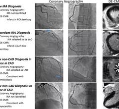
Cardiac MRI vs angiography for NSTEMI diagnosis. Cardiac MRI may better assess and treat patients who have experienced a type of heart attack caused by an extremely narrowed artery compared to standard angiography, reported a study published in Circulation. Overall, MRI led to a new diagnosis involving a damaged artery in 30% of patients, and a diagnosis unrelated to the artery in another 15%.

Is MRI better than angiography for diagnosing, treating certain heart attacks?

It can be hard for physicians to identify the damaged artery responsible for a non-ST segment elevation myocardial infarction (NSTEMI). Typically coronary angiography is the first choice, but is it the best?

May 30, 2019

Researchers develop new cardiac imaging tool to diagnose heart disease

The new method—cardiac functional MRI (cfMRI)—measures how the heart utilizes oxygen for both healthy patients and those with a form of heart disease.

May 30, 2019

Medical association releases consensus statement on intra-coronary imaging

The European Association of Percutaneous Cardiovascular Interventions (EAPCI) has published a clinical guidance statement regarding the use of intra-coronary imaging (IC), sharing the document during its EuroPCR Annual Meeting, May 21-24 in Paris.

May 22, 2019
heart.png

Radiologists performing fewer myocardial perfusion imaging exams

A recent decline in myocardial perfusion imaging (MPI) utilization has been attributed to legislative efforts, decreasing reimbursement and consolidation, but is that truly the case?

May 20, 2019

Around the web

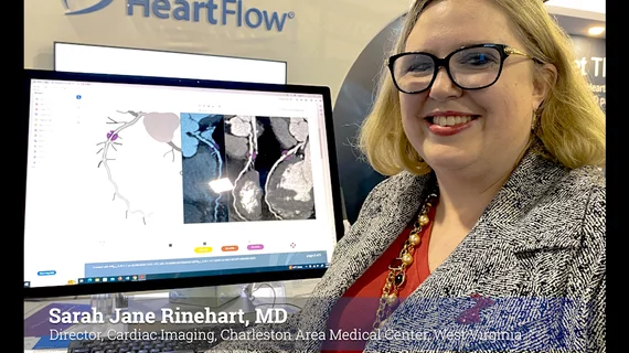
Automated AI-generated measurements combined with annotated CT images can improve treatment planning and help referring physicians and patients better understand their disease, explained Sarah Jane Rinehart, MD, director of cardiac imaging with Charleston Area Medical Center.

Two advanced algorithms—one for CAC scores and another for segmenting cardiac chamber volumes—outperformed radiologists when assessing low-dose chest CT scans. 

"Gen AI can help tackle repetitive tasks and provide insights into massive datasets, saving valuable time," Thomas Kurian, CEO of Google Cloud, said Tuesday. 

Trimed Popup
Trimed Popup