Cardiac Imaging

While cardiac ultrasound is the widely used imaging modality for heart assessments, computed tomography (CT), magnetic resonance imaging (MRI) and nuclear imaging are also used and are often complimentary, each offering specific details about the heart other modalities cannot. For this reason the clinical question being asked often determines the imaging test that will be used.

thi.d8d77f53.fill-1366x500-c0.format-jpeg.jpegquality-80.jpg

Musculoskeletal ultrasound may reduce unnecessary MRI, maintain accuracy

According to a study published in the Journal of the American College of Radiology, ultrasound may serve as an advanced imaging modality that reduces the need for additional musculoskeletal MRI examinations.

May 7, 2018

Document offers guidance on curbing radiation exposure during CV imaging

An expert consensus document published May 2 in the Journal of the American College of Cardiology offers recommendations for limiting radiation exposure to both patients and medical personnel during cardiovascular imaging procedures.

May 3, 2018

ACC releases strategies on reducing radiation from CV imaging

In recognition of the increased use of ionizing radiation-based cardiovascular imaging, the American College of Cardiology released a new expert document outlining best practices for minimizing exposure to patients and clinicians.

May 3, 2018
clinical-collaboration-platform.jpg

Premier Awards Carestream Health New Three-Year Agreement For Enterprise Image Management Solutions

Premier has awarded a three-year group purchasing agreement to Carestream Health for Enterprise Image Management Solutions.

May 2, 2018

Global longitudinal strain tops standard prediction method for heart failure

Global longitudinal strain (GLS), imaged via speckle-tracking, proved a superior predictor of mortality than left ventricular fraction (LVEF) in a study of acute heart failure patients, Cardiovascular Business reports.

May 2, 2018

3 key points on machine learning in cardiac CT

In light of the advances of machine learning (ML) in imaging, a team of researchers recently scoured ML-algorithm-based cardiovascular CT progress to offer an understanding of its benefits—and its limitations.

May 2, 2018
screen_shot_2018-05-01_at_11.29.17_am.png

Technologists can interpret chest x-rays as well as radiologists

With the appropriate amount of education and training, technologists can interpret chest x-rays with the same level of diagnostic accuracy and quality as radiologists, according to a study published in Academic Radiology.

May 1, 2018
James_Min

James K. Min named editor-in-chief of Journal of Cardiovascular Computed Tomography

James K. Min, MD, has been chosen to be the new editor-in-chief of the Journal of Cardiovascular Computed Tomography (JCCT), the official journal of the Society of Cardiovascular Computed Tomography (SCCT). He will serve a five-year term.
May 1, 2018

Around the web

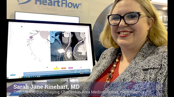
Automated AI-generated measurements combined with annotated CT images can improve treatment planning and help referring physicians and patients better understand their disease, explained Sarah Jane Rinehart, MD, director of cardiac imaging with Charleston Area Medical Center.

Two advanced algorithms—one for CAC scores and another for segmenting cardiac chamber volumes—outperformed radiologists when assessing low-dose chest CT scans. 

"Gen AI can help tackle repetitive tasks and provide insights into massive datasets, saving valuable time," Thomas Kurian, CEO of Google Cloud, said Tuesday. 

Trimed Popup
Trimed Popup