Computed Tomography

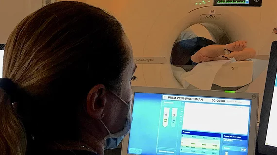
Computed tomography (CT) is a fast and accurate imaging modality often used in emergency settings and trauma imaging. CT scans, with or without (or both) iodinated contrast are frequently used to image the brain, chest, abdomen and pelvis, but also have post-imaging reconstructive capabilities for detailed orthopedic imaging. It is now a standard imaging modality in emergency rooms to quickly assess patients. CT uses a series of X-ray images shot as the gantry rotates around the patient. Computer technology assembles these into into a dataset volume than can be slices on any access, or advanced visualization software can extract specific parts of the anatomy for study. Find more content specific to cardiac CT.
Document offers guidance on curbing radiation exposure during CV imaging

An expert consensus document published May 2 in the Journal of the American College of Cardiology offers recommendations for limiting radiation exposure to both patients and medical personnel during cardiovascular imaging procedures.

May 3, 2018
James_Min

James K. Min named editor-in-chief of Journal of Cardiovascular Computed Tomography

James K. Min, MD, has been chosen to be the new editor-in-chief of the Journal of Cardiovascular Computed Tomography (JCCT), the official journal of the Society of Cardiovascular Computed Tomography (SCCT). He will serve a five-year term.
May 1, 2018
Massachusetts

Non-hospital-based facilities offering cardiac imaging services a rarity in Massachusetts

In 2017, Anthem implemented a new policy that requires outpatient MR and CT scans not considered medically necessary to be completed at a freestanding imaging facility to receive coverage. This went into effect in several states, sparking its fair share of controversy along the way, and a team of Boston-based researchers wanted to know how such a policy could impact patient care in Massachusetts.

March 5, 2018

Emergency CT for head trauma may be overused

New research suggests emergency patients are often given unwarranted CT scans to check for skull fractures and brain hemorrhage, resulting in wasted healthcare dollars and increasing exposure to radiation, according to an American Roentgen Ray Society (ARRS) press release.

February 26, 2018

Treatment costs of CT-related cancers could reach $2.6 billion over 10 years

Despite the many benefits of CT imaging—early diagnosis, enhanced diagnostic accuracy, improved outcomes, etc.—a small number of cancers resulting from patient exposure to radiation from CT scans can have a big financial impact on treatment costs, according to results of a study recently presented at the annual meeting of the American Roentgen Ray Society.

April 27, 2016

Budoff wins SCCT’s 2016 gold medal award

The Society of Cardiovascular Computed Tomography (SCCT) announced that Matthew J. Budoff, MD, had won its 2016 Gold Medal Award.

April 25, 2016
An ambulance-based mobile stroke unit created by the University of Texas Health Science Center at Houston (UTHealth) Medical School and Memorial Hermann-Texas Medical Center (TMC). The vehicle has a small head CT scanner to enable imm edit imaging to confirm if a patient had a stroke and what type before they arrive at the hospital to enable faster door to repercussion times, or faster door to tPA administration times. The unit also has a telemedicine system so a neurologist can evaluate patients remotely.

CT-based Mobile Stroke Care

Germany's ground-breaking use of CT scanners in ambulances to assess for possible stroke inspired one program in Houston to follow suit. Supporters predict it will transform care in the future. 

September 2, 2014
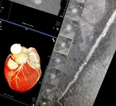
Example of a curved MPR image reconstruction of entire length of of a coronary artery on a cardiac CT scan to better show calcified and soft plaque burden inside the vessel. The thumbnail dots on the left side of the image are cross sectional views of the vessel. Siemens example on the expo floor.

The CCTA Playbook: A Guide to Coding, Reimbursement and Operations

A compendium of the business intelligence required to launch a CCTA service

March 2, 2009

Around the web

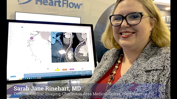
Automated AI-generated measurements combined with annotated CT images can improve treatment planning and help referring physicians and patients better understand their disease, explained Sarah Jane Rinehart, MD, director of cardiac imaging with Charleston Area Medical Center.

Two advanced algorithms—one for CAC scores and another for segmenting cardiac chamber volumes—outperformed radiologists when assessing low-dose chest CT scans. 

"Gen AI can help tackle repetitive tasks and provide insights into massive datasets, saving valuable time," Thomas Kurian, CEO of Google Cloud, said Tuesday. 

Trimed Popup
Trimed Popup