Nuclear Medicine

Nuclear medicine (also called molecular imaging) includes positron emission computed tomography (PET) and single photon emission computed tomography (SPECT) imaging. Nuclear imaging is achieved by injecting small amounts of radioactive material (radiopharmaceuticals) into patients before or during their scan. These can use sugars or chemical traits to bond to specific cells. The radioactive material is taken up by cells that consume the sugars. The radiation emitted from inside the body is detected by photon detectors outside the body. Computers take the data to assemble images of the radiation emissions. Nuclear images may appear fuzzy or ghostly rather than the sharper resolution from MRI and CT.  But, it provides metabolic information at a cellular level, showing if there are defects in the function of the heart, areas of very high metabolic activity associated with cancer cells, or areas of inflammation, data not available from other modalities. These noninvasive imaging exams are used to diagnose cancer, heart disease, Alzheimer’s and Parkinson’s disease, bone disorders and other disorders. 

MRI

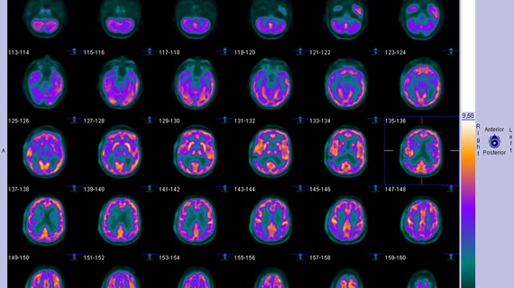
Radioactive substances unnecessary in new method for measuring brain glucose metabolism

Rather than administering radiolabeled glucose for exams, imagers give patients a small amount of a harmless glucose solution that is said to be equivalent to a can of a carbonated drink.

April 28, 2023
CT Scan

Accessibility, reimbursement and other issues limit uptake of PET/CT among oncologists, survey reveals

Difficulty obtaining PET/CT scans was reported by 55% of respondents, with 21% citing this as the greatest barrier in treating classic Hodgkin lymphoma.

April 27, 2023

MRI and PET findings could guide treatment of lingering concussion symptoms

Abnormalities of the thalamus could be to blame for persistent symptoms in the months following a concussion, according to new research.

April 26, 2023

Experts release new appropriate use criteria for lymphoscintigraphy in sentinel node mapping

The AUC includes a total of 32 clinical scenarios that have been grouped into four categories of cancer: breast, skin, cancer of other sites and lymphedema.

April 17, 2023
Alzheimer's

Imaging required to monitor effects of new Alzheimer's drug could inhibit its uptake

Leqembi can cause amyloid-related imaging abnormalities (ARIA), which must be assessed on imaging.

April 13, 2023
Tattoo

Imaging considerations included in tattoo guidelines for at-risk cancer population

Cases of mistaken malignancy have become more common as the popularity of tattoos has grown in recent years.

April 13, 2023
nuc med treatment for non-hodgkin lymphoma

New nuclear medicine treatment could potentially cure non-Hodgkin lymphoma

“If testing is successful in humans, this would represent an excellent new treatment option for patients with this disease.” 

April 11, 2023
PET/MRI of FAPI radiotracer uptake in Crohn's disease

New imaging technique could change how Crohn's disease is treated

The FAPI radiotracer experts used in this research can bind specifically to the connective tissue cells that cause intestinal wall fibrosis common to Crohn's disease.

April 5, 2023

Around the web

Automated AI-generated measurements combined with annotated CT images can improve treatment planning and help referring physicians and patients better understand their disease, explained Sarah Jane Rinehart, MD, director of cardiac imaging with Charleston Area Medical Center.

Two advanced algorithms—one for CAC scores and another for segmenting cardiac chamber volumes—outperformed radiologists when assessing low-dose chest CT scans. 

"Gen AI can help tackle repetitive tasks and provide insights into massive datasets, saving valuable time," Thomas Kurian, CEO of Google Cloud, said Tuesday. 

Trimed Popup
Trimed Popup