Structural Heart Disease

Structural heart diseases include any issues preventing normal cardiovascular function due to damage or alteration to the anatomical components of the heart. This is caused by aging, advanced atherosclerosis, calcification, tissue degeneration, congenital heart defects and heart failure. The most commonly treated areas are the heart valves, in particular the mitral and aortic valves. These can be replaced through open heart surgery or using cath lab-based transcatheter valves or repairs to eliminate regurgitation due to faulty valve leaflets. This includes transcatheter aortic valve replacement (TAVR). Other common procedures include left atrial appendage (LAA) occlusion and closing congenital holes in the heart, such as PFO and ASD. A growing area includes transcatheter mitral repair or replacement and transcatheter tricuspid valve repair and replacement.
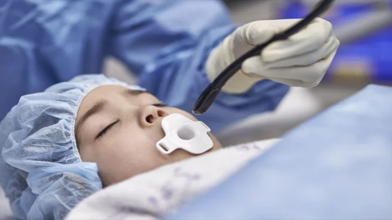
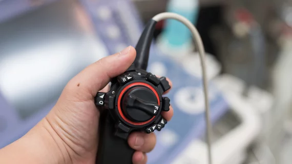
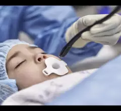
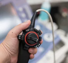
Philips received U.S. Food and Drug Administration (FDA) clearance for a compact transesophageal echocardiography (TEE) ultrasound transducer. The X11-4t Mini 3D TEE transducer was designed to improve image quality when evaluating certain patient populations, including pediatric patients and adults who present with a heightened risk of complications.

FDA clears smaller 3D TEE transducer for imaging children, high-risk adult patients

The newly approved device is much smaller than previous offerings, helping operators capture 3D images during structural heart evaluations. 

January 31, 2024
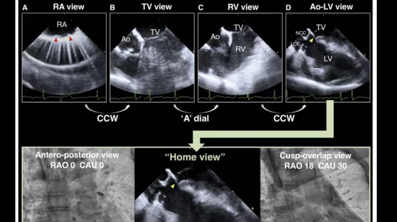
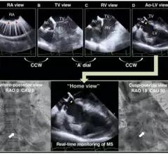
Self-expandable TAVR cusp-overlap ICE

ICE guidance reduces risk of permanent pacemaker implantation after TAVR with a self-expandable valve

Heart teams can limit the risk of conduction disturbances that lead to permanent pacemaker implantation by utilizing both the cusp-overlap method and intracardiac echocardiography.

January 17, 2024
transesophageal echocardiography (TEE)

New expert guidelines: Start PHV evaluations with echocardiography, but other imaging modalities can provide value

New guidelines from the American Society of Echocardiography, made in collaboration with the Society for Cardiovascular Magnetic Resonance and Society of Cardiovascular Computed Tomography, represent an update of the group's original recommendations from 2009.

January 9, 2024
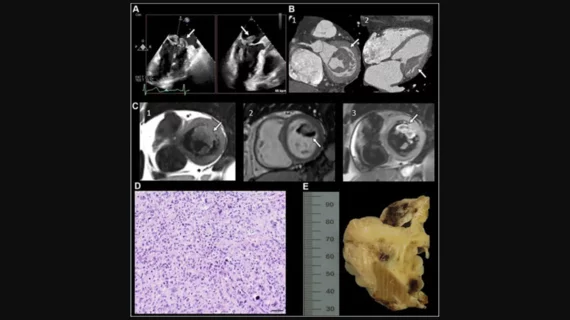
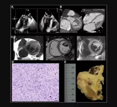
“Undifferentiated Cardiac Sarcoma of the Mitral Valve: Multimodal Imaging Assessment" Radiology: Cardiothoracic Imaging RSNA

Radiologists select 2023's top images in cardiothoracic imaging

“Cardiothoracic imaging continues to evolve, fueled by technical innovations such as dynamic chest radiography, digital tomosynthesis and dark-field radiography," one radiologist said.

December 21, 2023
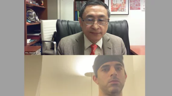
Arzhang Fallahi, MD, and David Hsi, MD, discussing imaging-based aortic stenosis screening

Q&A: Cardiologists explore the potential impact of a screening program for aortic stenosis

We already screen patients for breast cancer and lung cancer on a regular basis. Why not establish screening programs for aortic stenosis?

September 11, 2023
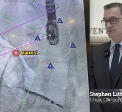
Video of ASE President Stephen Little, MD, discussing trends in interventional echo at the ASE 2023 annual meeting.

Interventional echocardiography expected to grow thanks to new structural heart procedures

American Society of Echocardiography (ASE) President Stephen Little, MD, says there will be an increasing need for interventional echocardiographers in the near future.

August 22, 2023
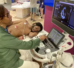
Patient being scanned on Philips Epiq ultrasound cardiac echo ASE 2023.

PHOTO GALLERY: The trends and technologies at ASE 2023

Browse through some of the many highlights from the American Society of Echocardiography's 2023 annual meeting. 

July 7, 2023
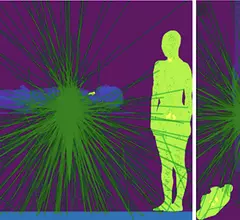
X-ray photon trajectory during the simulation phantom study from the side and top views. Due to scattering of the X-rays when they hit the lower end of the patient bed, exposure in mainly to the lower body of the interventional echocardiographer performing transesophageal echocardiography. The green lines are the scattered photon trajectories calculated by Monte Carlo simulation in the study.

Radiation exposure in the cath lab: Tracking the impact on interventional echocardiographers

Researchers found that echocardiographers in the cath lab are exposed to high doses of radiation on the right half of their body, especially the waist and lower body. 

April 28, 2023

Around the web

Automated AI-generated measurements combined with annotated CT images can improve treatment planning and help referring physicians and patients better understand their disease, explained Sarah Jane Rinehart, MD, director of cardiac imaging with Charleston Area Medical Center.

Two advanced algorithms—one for CAC scores and another for segmenting cardiac chamber volumes—outperformed radiologists when assessing low-dose chest CT scans. 

"Gen AI can help tackle repetitive tasks and provide insights into massive datasets, saving valuable time," Thomas Kurian, CEO of Google Cloud, said Tuesday. 

Trimed Popup
Trimed Popup