Structural Heart Disease

Structural heart diseases include any issues preventing normal cardiovascular function due to damage or alteration to the anatomical components of the heart. This is caused by aging, advanced atherosclerosis, calcification, tissue degeneration, congenital heart defects and heart failure. The most commonly treated areas are the heart valves, in particular the mitral and aortic valves. These can be replaced through open heart surgery or using cath lab-based transcatheter valves or repairs to eliminate regurgitation due to faulty valve leaflets. This includes transcatheter aortic valve replacement (TAVR). Other common procedures include left atrial appendage (LAA) occlusion and closing congenital holes in the heart, such as PFO and ASD. A growing area includes transcatheter mitral repair or replacement and transcatheter tricuspid valve repair and replacement.
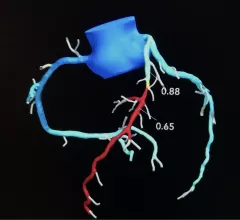
Performing CT-derived fractional flow reserve (CT-FFR) before transcatheter aortic valve replacement (TAVR) improves the accuracy of coronary CT angiography (CCTA) and helps limit unneeded invasive coronary angiography (ICA), according to a new study published in JACC: Cardiovascular Interventions. Heartflow

CT-FFR before TAVR improves detection of coronary artery disease, limits invasive imaging exams

CT-FFR, which recently got a boost from the 2021 AHA/ACC chest pain guidelines, could play a key role for clinicians hoping to screen TAVR patients for coronary heart disease. 

May 11, 2022
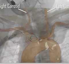
transcatheter aortic valve replacement (TAVR) procedure guided by CT fusion imaging on a patient with heart failure and a blood clot. CT fusion imaging-guided deployment of the Sentinel Cerebral Protection System during a TAVR procedure. Image courtesy of Vasudev et al.

Cardiologists and surgeons perform first TAVR using CT imaging guidance on HF patient with blood clot

“We believe this case is potentially groundbreaking," one interventional cardiologist said. 

May 6, 2022

Around the web

Automated AI-generated measurements combined with annotated CT images can improve treatment planning and help referring physicians and patients better understand their disease, explained Sarah Jane Rinehart, MD, director of cardiac imaging with Charleston Area Medical Center.

Two advanced algorithms—one for CAC scores and another for segmenting cardiac chamber volumes—outperformed radiologists when assessing low-dose chest CT scans. 

"Gen AI can help tackle repetitive tasks and provide insights into massive datasets, saving valuable time," Thomas Kurian, CEO of Google Cloud, said Tuesday. 

Trimed Popup
Trimed Popup