Structural Heart Disease

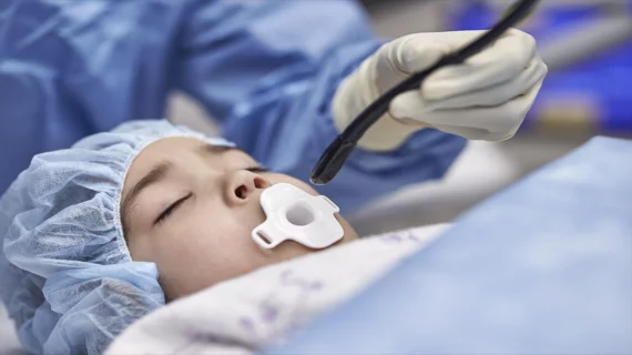
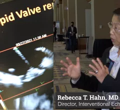
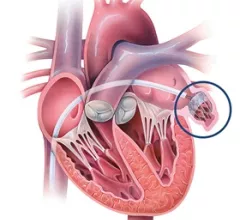
Structural heart diseases include any issues preventing normal cardiovascular function due to damage or alteration to the anatomical components of the heart. This is caused by aging, advanced atherosclerosis, calcification, tissue degeneration, congenital heart defects and heart failure. The most commonly treated areas are the heart valves, in particular the mitral and aortic valves. These can be replaced through open heart surgery or using cath lab-based transcatheter valves or repairs to eliminate regurgitation due to faulty valve leaflets. This includes transcatheter aortic valve replacement (TAVR). Other common procedures include left atrial appendage (LAA) occlusion and closing congenital holes in the heart, such as PFO and ASD. A growing area includes transcatheter mitral repair or replacement and transcatheter tricuspid valve repair and replacement.

Example of interventional echocardiography TEE imaging superimposed on live fluoro during a transseptal puncture for a MitraClip procedure.

New ASE guideline outlines training standards for interventional echocardiography

The American Society of Echocardiography (ASE) released a new guideline document that outlines uniform training standards for interventional sonographers guiding structural heart procedures.

April 26, 2023
Brian Lindman, MD, MSCI, medical director of the Structural Heart and Valve Center and an associate professor of medicine, Vanderbilt University Medical Center, explains the new American Heart Association (AHA) Target Aortic Stenosis (AS) program. It was announced at the AHA 2022 meeting and is designed to help screen with echo to find more AS patients.

VIDEO: AHA says aortic stenosis is undertreated and underdiagnosed

Brian Lindman, MD, medical director of the Structural Heart and Valve Center, Vanderbilt University Medical Center, explains the new American Heart Association (AHA) Target Aortic Stenosis (AS) program to screen with echo to find more AS patients. 

November 14, 2022
mixed reality LAAO Chase Western Reserve MRI. The group—which includes engineers, cardiologist, radiologists and other specialists—will attempt to perform a robotic-controlled left atrial appendage occlusion (LAAO) on a patient inside an MRI scanner.

Researchers receive $3.7M to attempt robotic heart surgery inside MRI scanner

The group—which includes engineers, cardiologist, radiologists and other specialists—will attempt to perform a robotic-controlled left atrial appendage occlusion (LAAO) on a patient inside an MRI scanner.

October 20, 2022
CT has been used for planning and procedural guidance in pulmonary vein isolation (PVI) for about 20 years. It shows the anatomy to the electrophysiologist. The EPs also can load the imaging into their EP mapping system. It also shows them the location of the esophagus and the phrenic nerve so they can avoid these during the procedures. #PVI #EPeeps

VIDEO: The expanding role of cardiac CT in electrophysiology

Harold Litt, MD, division chief of cardiothoracic imaging and director of the Center for Advanced CT Imaging Sciences at the Perelman School of Medicine of the University of Pennsylvania, explains the role of cardiac CT in EP procedures.

October 12, 2022
CT imaging showing the congenital heart defect of left heart hypoplastic syndrome.

VIDEO: Congenital heart imaging with cardiac CT

As the population of patients with adult congenital heart disease grows, they are presenting to adult cardiology clinics and being imaged with CT. Many also do not have access or cannot be imaged by MRI, said Renee Bullock-Palmer, MD.

September 28, 2022
Examples of structural heart transcatheter valve replacement procedure planning CT scans and post procedure followup for TMVR and TAVR.

VIDEO: CT imaging for TAVR and TMVR structural heart interventions

Joao Cavalcante, MD, director, cardiac MRI and structural CT labs, Minneapolis Heart Institute, discusses the use of cardiac CT imaging to plan and guide structural heart procedures. 

September 15, 2022
Interview with Rebecca T. Hahn, MD, Professor of Medicine at Columbia University Irving Medical Center, Chief Scientific Officer of the Echo Core Lab at the Cardiovascular Research Foundation and Director of Interventional Echocardiography at the Columbia Structural Heart and Valve Center. She discusses some of the trends of growing use of interventional echocardiographic guidance in transcatheter structural heart procedures, the growing number of tricuspid valve procedures, and use of 3D ICE.

VIDEO: Trends in structural heart procedural imaging - a discussion with Rebecca Hahn

Rebecca T. Hahn, MD, Director of Interventional Echocardiography at the Columbia Structural Heart and Valve Center, discusses some of the trends in the growing use of interventional echocardiographic guidance in transcatheter structural heart procedures.

September 9, 2022
Illustration of a left atrial appendage occlusion (LAAO) procedure using a Watchman device. Image courtesy of Johns Hopkins Medicine. #LAA #LAAO

Pre-procedure CT imaging benefits LAA occlusion in Henry Ford study

There is not agreement among the top structural heart experts if CT is required for left atrial appendage occlusion (LAAO), but a Henry Ford Hospital study shows it can improve outcomes.

August 5, 2022

Around the web

Automated AI-generated measurements combined with annotated CT images can improve treatment planning and help referring physicians and patients better understand their disease, explained Sarah Jane Rinehart, MD, director of cardiac imaging with Charleston Area Medical Center.

Two advanced algorithms—one for CAC scores and another for segmenting cardiac chamber volumes—outperformed radiologists when assessing low-dose chest CT scans. 

"Gen AI can help tackle repetitive tasks and provide insights into massive datasets, saving valuable time," Thomas Kurian, CEO of Google Cloud, said Tuesday. 

Trimed Popup
Trimed Popup