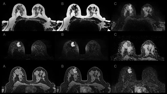
A. Axial contrast-enhanced CT image shows 1.7 cm lesion (arrow) with ill-defined margin at left side of superior mesenteric artery. B. On axial T2-weighted image, mass is hyperintense to paravertebral muscle (arrow). C. On arterial phase post-contrast T1-weighted image, mass shows mild enhancement (arrow) compared to unenhanced image; enhancement pattern classified as early. D. On DWI with b-value of 800 mm2/s, mass is hyperintense (arrow) to paravertebral muscle. E. On ADC map, mass is visually hypointense (arrow) to paravertebral muscle. Findings indicate presence of qualitative diffusion restriction; lesion ADC value was 0.96 × 10⁻³ mm2/s. F. Axial image from CT performed 6 months after MRI shows increase in lesion’s greatest axial diameter by more than 5 mm (arrow), indicating soft tissue abnormality represents locally recurrent tumor.
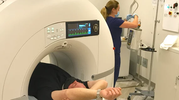
Photo and caption courtesy of American Journal of Roentgenology (AJR), American Roentgen Ray Society (ARRS). Used with permission.


, American Roentgen Ray Society (ARRS). Used with permission.")