Structured communication feedback systems between radiologists and intensive care unit physicians can significantly reduce instances of adverse events.
Dynamic chest radiography was recently shown to be comparable to lung ventilation-perfusion scanning for detecting chronic thromboembolic pulmonary hypertension.
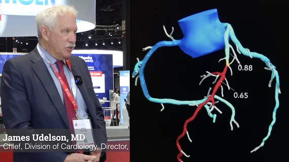
James Udelson, MD, an investigator involved with the PRECISE trial, explained how patient outcomes were improved by 70% from the current standard of care.
While electronically cropping an image may seem like a harmless act, the habit is not without unintended consequences, the authors of a newpaper recently explained.
In the case of a convicted murderer, a New York neuro specialist cited a slew of neuroimaging findings that indicate “severe dysfunction” that could have numbed the defendant’s “brakes of inhibition.”
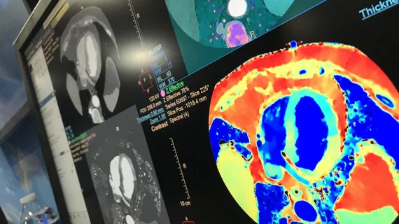
Suhny Abbara, MD, editor of Radiology: Cardiothoracic Imaging and chief of cardiothoracic imaging for University of Texas Southwestern Medical Center, discusses how spectral computed tomography (CT) can help both cardiac and general CT imaging.
New research indicates that there is significant reader variability in COVID classifications among different specialties when chest X-rays alone are the diagnostic tool of choice.
The radiologist involved in the incident indicated that the department’s burdensome workload resulted in reduced reading times, saying that this made their departmental standard of double reading scans “virtually unattainable."