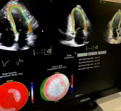
Echocardiography

Cardiac ultrasound uses reflected sound waves (echos) to create images of anatomy inside the body. Echocardiograms are the primary cardiac imaging modality used to assess the heart and diagnose or track cardiac issues. Echo is the gold standard imaging modality to assess the heart, particularly with calculating left ventricular ejection fraction (LVEF), which is a measure of cardiac output. In addition to noninvasive standard transthoracic echo (TTE), invasive transesophgeal echo (TEE) is also used when clearer, more detailed imaging of the heart is needed. Both 3D and 4D echo echo systems are rapidly gaining wider adoption and enable new types of assessments, especially in the structural heart space and in transcatheter procedural guidance. Find news on general ultrasound imaging.

Seeing the Big Picture: Training Today’s Imagers to ‘Think Multimodality’

Cardiologists are receiving more exposure to different imaging modalities during their fellowships, but their job prospects and training vary widely. A more comprehensive and multimodality training approach could lead to better results.  

January 18, 2018

Genetic variants could be key to identifying chemo-induced cardiotoxicity

As life expectancy continues to expand for cancer patients, clinicians are increasingly dealing with oncological complications like cardiotoxicity, according to a medical team in the Netherlands—and those doctors are met with a paucity of research on the topic.

January 15, 2018

Automated analysis of 3D echocardiography shows potential in clinical practice

Automated 3D echocardiography (3DE) analysis using a new, commercially available algorithm has allowed University of Chicago researchers to accurately quantify left-heart size and function in two-thirds of 300 consecutive patients. They conclude the technology can be useful in clinical practice despite its known workflow-interruptive drawbacks—especially when the echocardiographer has the know-how to correct for its shortcomings.

July 10, 2017
Geoffrey A. Rose, MD, President, Sanger Heart & Vascular Institute

Pathways for Quality Care: Echocardiographers Ponder a Playbook for Imaging in Myocardial Infarction

Physicians in fields like cardiology have traditionally looked to clinical practice guidelines to help articulate the best evidence-based care for patients. The rapidly growing movement to value-based care is prompting clinicians—including echocardiographers—to carefully weigh a more focused and integrative approach to delivering consistent, quality medicine: care pathways.

April 21, 2017
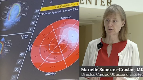
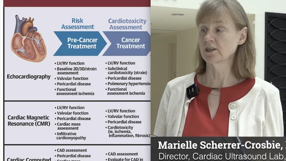
To prevent the risk of patients developing heart failure from chemotherapy agents in cancer care, patients are typically have their left ventricular ejection fraction (LVEF) or myocardial strain monitored using either echocardiography or equilibrium radionuclide angiography/multigated acquisition (ERNA/MUGA). If cardiac damager occurs, the treatment is discontinue or pause treatment. Cardiac imaging to assess chemotherapy-induced cardiotoxicity using strain echo.

Succeeding with Cancer: Using Imaging to Avoid Treatment-induced Heart Failure

Treating today’s cancer patient no longer means simply targeting the cancer. Given the known cardiotoxicities of some established chemotherapies and the possibility that newer approaches may damage the heart, oncologists, cardiologists and imaging specialists now work together to detect and minimize the risk of treatment-induced heart failure.

March 22, 2017
Sharon L. Mulvagh, MD

Goal Reversal? Study Suggests Echocardiography Could Have an Underuse Problem

ASE says new data may be a “wake-up call” to recognize the value of echocardiography.  

May 26, 2016

ASE, GE partner in India for cardiovascular ultrasound training

The American Society of Echocardiography (ASE) and GE Healthcare have teamed to provide a cardiovascular ultrasound training event for healthcare providers caring for underserved populations in rural northwest India.

December 10, 2012

ASE: Are cardiovascular sonographers at high risk of radiation exposure?

There is growing concern among cardiac sonographers about the amount of radiation exposure they experience on the job and few say their workplace has a formal policy to address radiation safety for sonographers, according to a survey conducted by the American Society of Echocardiography.

September 30, 2011

Around the web

Automated AI-generated measurements combined with annotated CT images can improve treatment planning and help referring physicians and patients better understand their disease, explained Sarah Jane Rinehart, MD, director of cardiac imaging with Charleston Area Medical Center.

Two advanced algorithms—one for CAC scores and another for segmenting cardiac chamber volumes—outperformed radiologists when assessing low-dose chest CT scans. 

"Gen AI can help tackle repetitive tasks and provide insights into massive datasets, saving valuable time," Thomas Kurian, CEO of Google Cloud, said Tuesday. 

Trimed Popup
Trimed Popup