Echocardiography

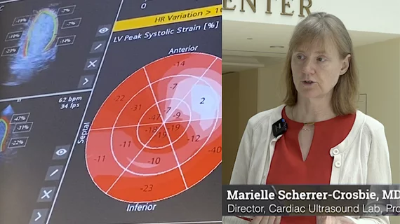
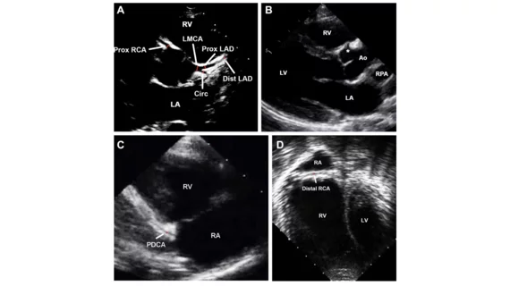
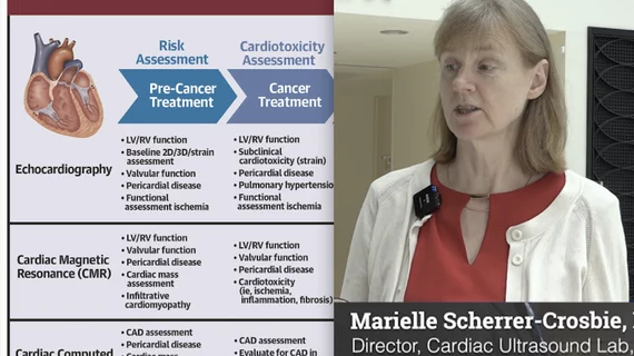
Cardiac ultrasound uses reflected sound waves (echos) to create images of anatomy inside the body. Echocardiograms are the primary cardiac imaging modality used to assess the heart and diagnose or track cardiac issues. Echo is the gold standard imaging modality to assess the heart, particularly with calculating left ventricular ejection fraction (LVEF), which is a measure of cardiac output. In addition to noninvasive standard transthoracic echo (TTE), invasive transesophgeal echo (TEE) is also used when clearer, more detailed imaging of the heart is needed. Both 3D and 4D echo echo systems are rapidly gaining wider adoption and enable new types of assessments, especially in the structural heart space and in transcatheter procedural guidance. Find news on general ultrasound imaging.

Elastography, echogenicity discriminate plaques in symptomatic stenosis patients

Combining ultrasound elastography and echogenicity analysis may better distinguish between plaques in patients with symptomatic internal carotid artery (ICA) stenosis than in those without symptoms, according to an Aug. 30 study in the American Journal of Roentgenology.

September 4, 2018
Cardiovascular

Left ventricle-only echocardiography saves time, resources

A group of researchers from a Veterans Affairs health system in California shared their experience with limited left ventricular echocardiography, something they believe could reduce the cost and time required for testing in select patients.

August 3, 2018

Left ventricle-focused echocardiography quick, cost-effective

A group of California researchers believes a limited transthoracic echocardiogram (TTE) that solely evaluates the left ventricle could be more efficient and cost-effective in certain patients compared to whole echocardiography.

July 31, 2018
Jonathan Lindner

Jonathan Lindner elected president of the American Society of Echocardiography

The 17,000-member American Society of Echocardiography (ASE) named Jonathan R. Lindner, MD, president of the organization during its 29th annual scientific sessions in Nashville, Tennessee.

June 27, 2018

FDA-approved AI echocardiogram software bests cardiologists in reducing LVEF variability

A deep-learning software that can automatically calculate left ventricular ejection fraction (LVEF) with less variability than a cardiologist recently received approval from the U.S. Food and Drug Administration (FDA).

June 26, 2018
Cancer overdiagnosis

Is coronary CTA or stress echocardiography better for chest pain triage?

Coronary computed tomography angiography (CTA) is the current go-to for triaging chest pain patients in the emergency department (ED), but a recent study found the often-overlooked stress echocardiography (SE) may actually be safer and provide patients with a better overall experience.

June 15, 2018

Data fatigue: Machine learning predicts post-echocardiography survival with EHR assistance

Machine learning can accurately predict survival after echocardiography by analyzing unique data produced from heart images and electronic health record (EHR) information, according to a recent study in the Journal of the American College of Cardiology: Cardiovascular Imaging.

June 15, 2018

New registry seeks to improve cardiovascular ultrasounds

ImageGuideEcho—the first registry devoted solely to measuring quality in cardiovascular ultrasounds— is now open to U.S. physicians.

January 29, 2018

Around the web

Automated AI-generated measurements combined with annotated CT images can improve treatment planning and help referring physicians and patients better understand their disease, explained Sarah Jane Rinehart, MD, director of cardiac imaging with Charleston Area Medical Center.

Two advanced algorithms—one for CAC scores and another for segmenting cardiac chamber volumes—outperformed radiologists when assessing low-dose chest CT scans. 

"Gen AI can help tackle repetitive tasks and provide insights into massive datasets, saving valuable time," Thomas Kurian, CEO of Google Cloud, said Tuesday. 

Trimed Popup
Trimed Popup