Cardiac Imaging

While cardiac ultrasound is the widely used imaging modality for heart assessments, computed tomography (CT), magnetic resonance imaging (MRI) and nuclear imaging are also used and are often complimentary, each offering specific details about the heart other modalities cannot. For this reason the clinical question being asked often determines the imaging test that will be used.
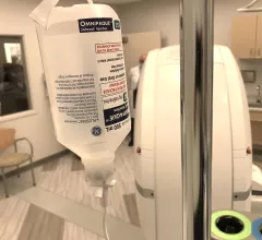
With the shortage, which pertains specifically to all formulations and concentrations of GE Healthcare’s Omnipaque (iohexol), expected to last until mid-June, several organizations have eluded that conservation efforts are of critical importance.#contrastshortage #CTcontrast #iodinatedcontrast

GE provides update on contrast media shortage

The COVID lockdowns at the Shanghai plant began on March 31, and although the plant there has since resumed operations, they are not yet at 100% capacity.

May 18, 2022

VIDEO: Cardiology getting more involved in pulmonary embolism response teams

Terry Bowers, MD, director of vascular medicine at Beaumont Hospital and national co-chair of the Pulmonary Embolism Research Collaborative (PERC), explains the trend toward creating pulmonary embolism (PE) response teams that include cardiology. 

May 18, 2022
A comparison on the black hole at the center of the Milky Way galaxy (left) with PET cardiac myocardial perfusion nuclear imaging exams (right).  The cardiac diagnosis of the galactic heart is motion artifact. Black hole image from the EHT Collaboration

Cardiologist and ASNC president diagnoses the heart of the galaxy based on black hole imaging

The first image of the black hole at the heart of our Milky Way galaxy amazed the scientific community, but left cardiologists with questions about the true cardiac health of the galaxy.

May 16, 2022
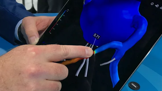
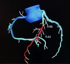
Performing CT-derived fractional flow reserve (CT-FFR) before transcatheter aortic valve replacement (TAVR) improves the accuracy of coronary CT angiography (CCTA) and helps limit unneeded invasive coronary angiography (ICA), according to a new study published in JACC: Cardiovascular Interventions. Heartflow

CT-FFR before TAVR improves detection of coronary artery disease, limits invasive imaging exams

CT-FFR, which recently got a boost from the 2021 AHA/ACC chest pain guidelines, could play a key role for clinicians hoping to screen TAVR patients for coronary heart disease. 

May 11, 2022
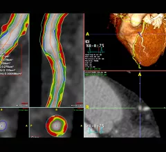
An example of CT imaging coronary plaque assessment on TeraRecon's advanced visualization software.

VIDEO: Use of CT to assess coronary plaques

Leslee Shaw, PhD, director of The Blavatnik Family Women’s Health Research Institute, Mount Sinai Hospital, New York, explains how cardiac computed tomography (CT) can be used to assess coronary plaques.

May 9, 2022
Patients with heart failure with preserved ejection fraction (HFpEF), who face a high mortality risk and do not respond to conventional therapies, are changing the way clinicians think about heart failure.

Is coronary heart disease on CT associated with early development of COPD?

"This awareness is important to avoid oversight of symptoms like dyspnea and vague chest discomfort, which can easily be interpreted as symptoms caused by the known disease COPD,” experts involved in the study said.

May 9, 2022
transcatheter aortic valve replacement (TAVR) procedure guided by CT fusion imaging on a patient with heart failure and a blood clot. CT fusion imaging-guided deployment of the Sentinel Cerebral Protection System during a TAVR procedure. Image courtesy of Vasudev et al.

Cardiologists and surgeons perform first TAVR using CT imaging guidance on HF patient with blood clot

“We believe this case is potentially groundbreaking," one interventional cardiologist said. 

May 6, 2022
A new artificial intelligence tool can detect heart disease on cardiovascular MRI scans in seconds with equal or superior precision as clinicians. 

Could cardiac MRI become the reference standard for diagnosing heart failure?

Cardiac MRI may offer a more accurate and less invasive method for diagnosing heart failure, a new study published in the European Heart Journal shows. 

May 5, 2022

Around the web

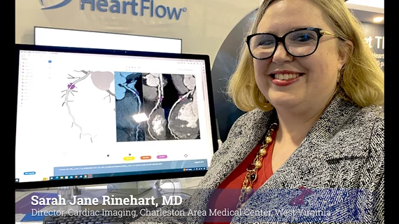
Automated AI-generated measurements combined with annotated CT images can improve treatment planning and help referring physicians and patients better understand their disease, explained Sarah Jane Rinehart, MD, director of cardiac imaging with Charleston Area Medical Center.

Two advanced algorithms—one for CAC scores and another for segmenting cardiac chamber volumes—outperformed radiologists when assessing low-dose chest CT scans. 

"Gen AI can help tackle repetitive tasks and provide insights into massive datasets, saving valuable time," Thomas Kurian, CEO of Google Cloud, said Tuesday. 

Trimed Popup
Trimed Popup