Molecular Imaging

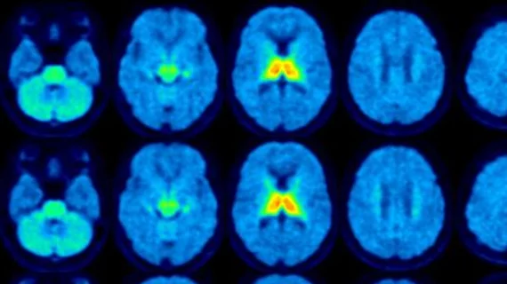
Molecular imaging (also called nuclear medicine or nuclear imaging) can image the function of cells inside the body at the molecular level. This includes the imaging modalities of positron emission computed tomography (PET) and single photon emission computed tomography (SPECT) imaging. How does PET and SPECT imaging work? Small amounts of radioactive material (radiopharmaceuticals) injected into a patient. These can use sugars or chemical traits to bond to specific cells. The radioactive material is taken up by cells that consume the sugars. The radiation emitted from inside the body is detected by photon detectors outside the body. Computers take the data to assemble images of the radiation emissions. Nuclear images may appear fuzzy or ghostly rather than the sharper resolution from MRI and CT.  But, it provides metabolic information at a cellular level, showing if there are defects in the function of the heart, areas of very high metabolic activity associated with cancer cells, or areas of inflammation, data not available from other modalities. These noninvasive imaging exams are used to diagnose cancer, heart disease, Alzheimer’s and Parkinson’s disease, bone disorders and other disorders. 

Molecular breast cancer imaging may be called on to reduce overdiagnosis

This issue isn't about false positives, it is about subjecting patients to treatment after mammography finds a small lesion, but when in all actuality that spot on the scan poses no real threat to the patient. Some cancers are never progressive and do not require serious treatment when to do so would come with its own risks and potential complications.

April 15, 2014

ABT implements first Dose on Demand Biomarker Generator in Russia

ABT Molecular Imaging has successfully implemented the first BG-75 Biomarker Generator in Russia. The project was completed in cooperation with the Diagnostic and Treatment Center of the International Institute of Biological Systems (DTC IIBS), St. Petersburg, Russia, and its U.S. representative, Owen Kane Molecular Imaging, Inc. (Owen Kane). 

April 15, 2014

The rising tide of Alzheimer’s

As the baby boomers shift into retirement, it’s always been known that the prevalence of age-related conditions such as Alzheimer’s disease was going to increase. Numbers released last month from the Alzheimer’s Association provided the latest estimate of just how severe the impact will be.

April 10, 2014

EU aims for clinical research transparency

Drug policy in the European Union could change in the not too distant future depending on a vote regarding a law proposed to discourage pharmaceutical companies from playing favorites when it comes to clinical trials.

April 7, 2014

New MRI technique tracks gene to help explain formation of memories

Biological engineers at the Massachusetts Institute of Technology have developed a method to adapt MRI to visualize gene activity in an effort to better understand the processes behind the formation of memories.

March 26, 2014

UltraSPECT and ASNC partner to provide technologist travel awards to attend ASNC2014

UltraSPECT, provider of the most cost-effective solutions for meeting American Society of Nuclear Cardiology (ASNC) guidelines for low dose nuclear medicine imaging, announces today its support of nuclear medicine technologists via six new grants.
March 6, 2014

Lab on the field: Combining molecular imaging and microfluidics

A tiny molecular imaging system could be used on the field to measure brain injury or in the lab to capture individual molecules that betray the biology of cancer, neurodegenerative diseases such as Alzheimer's and viruses like human immunodeficiency virus (HIV). 

January 27, 2014

Sodium fluoride PET/CT points to plaques, predicts heart attack

A PET/CT study followed 40 sufferers of heart attacks and just as many matched controls imaged with conventional coronary angiography and sodium fluoride PET/CT to gauge vulnerable plaques and see potential risk of heart attack.

January 22, 2014

Around the web

Automated AI-generated measurements combined with annotated CT images can improve treatment planning and help referring physicians and patients better understand their disease, explained Sarah Jane Rinehart, MD, director of cardiac imaging with Charleston Area Medical Center.

Two advanced algorithms—one for CAC scores and another for segmenting cardiac chamber volumes—outperformed radiologists when assessing low-dose chest CT scans. 

"Gen AI can help tackle repetitive tasks and provide insights into massive datasets, saving valuable time," Thomas Kurian, CEO of Google Cloud, said Tuesday. 

Trimed Popup
Trimed Popup