Molecular Imaging

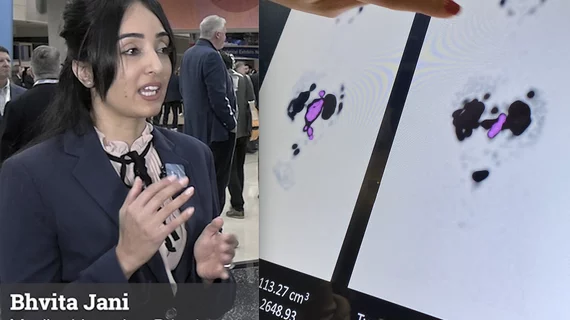
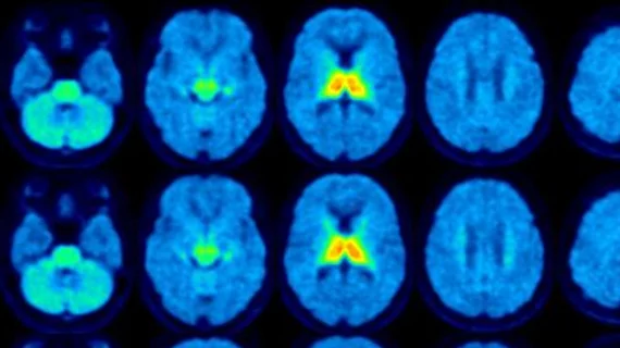
Molecular imaging (also called nuclear medicine or nuclear imaging) can image the function of cells inside the body at the molecular level. This includes the imaging modalities of positron emission computed tomography (PET) and single photon emission computed tomography (SPECT) imaging. How does PET and SPECT imaging work? Small amounts of radioactive material (radiopharmaceuticals) injected into a patient. These can use sugars or chemical traits to bond to specific cells. The radioactive material is taken up by cells that consume the sugars. The radiation emitted from inside the body is detected by photon detectors outside the body. Computers take the data to assemble images of the radiation emissions. Nuclear images may appear fuzzy or ghostly rather than the sharper resolution from MRI and CT.  But, it provides metabolic information at a cellular level, showing if there are defects in the function of the heart, areas of very high metabolic activity associated with cancer cells, or areas of inflammation, data not available from other modalities. These noninvasive imaging exams are used to diagnose cancer, heart disease, Alzheimer’s and Parkinson’s disease, bone disorders and other disorders. 

Advances in genetics and medical imaging meet Richard III

Shakespeare called King Richard III a hunchback, but new genetic and reconstructive CT data show this story had more of a twist. Until recently, the exact nature and whereabouts of Richard's physical deformity was a mystery. While questions still daunt researchers, a compelling picture of the monarch's spine has emerged.

May 30, 2014

Biobank launches brain study

In this video, BBC reporter Fergus Walsh is the first to undergo an MRI scan as part of the largest scan study to date and describes how the study is evolving.

May 23, 2014

IBA Molecular awarded 3-year group purchasing agreement with Premier, Inc.

IBA Molecular North America, Inc. (IBA Molecular) announced the signing of a 3-year group purchasing agreement with Premier, Inc. (Premier), a leading health care improvement company, to offer PET radiopharmaceuticals, including 18F-Fluorodeoxyglucose (FDG), a diagnostic radiotracer used in oncology, neurology and cardiology, to Premier member imaging centers across the country.

May 23, 2014

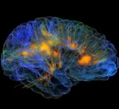
Glass Brain shows 3D detail of brain connectivity

Watch a fly-through of Glass Brain, a 3D imagining of the human brain by researchers at the University of California San Francisco. This video shows the 3D brain model made possible by electroencephalography data and white matter tract reconstruction from MRI diffusion tensor imaging. UCSF is using the technology to research possible treatments for neurodegenerative disease.

May 16, 2014

Revisiting the human genome

The human genome was first mapped just a little over a decade ago. Its 3 billion bits of DNA are the pages of the human instruction manual. Read this great review featuring Eric S. Lander from MIT, Harvard, and the Broad Institute in Cambridge to see how far we've come and what the future of genomics holds.

May 15, 2014

Clarifying cancer with 3D modeling

The shape of cancer genomics may be a new calling card for 3D oncologic imaging. Studying the formation of cancer cell genomes could potentially provide more information about a patient's cancer than just focusing on genetic expression. Scientists at McGill University in Montreal are finding that the form of a cancer cell genome can even tell them what subtype of cancer a patient has.

May 15, 2014

Bristol Myers Squibb nabs iPierian in $175M buyout

Biopharmaceutical giant Bristol-Myers Squibb now holds the power on a pipeline of neurodegenerative disease therapies after buying San Francisco-based iPierian for $175 million.

May 2, 2014

Genetic testing developed to end era of brain disorder in Turkey

Advanced gene sequencing has found a single genetic mutation essential for a brain disorder that has been plaguing Turkish families for centuries.

April 30, 2014

Around the web

Automated AI-generated measurements combined with annotated CT images can improve treatment planning and help referring physicians and patients better understand their disease, explained Sarah Jane Rinehart, MD, director of cardiac imaging with Charleston Area Medical Center.

Two advanced algorithms—one for CAC scores and another for segmenting cardiac chamber volumes—outperformed radiologists when assessing low-dose chest CT scans. 

"Gen AI can help tackle repetitive tasks and provide insights into massive datasets, saving valuable time," Thomas Kurian, CEO of Google Cloud, said Tuesday. 

Trimed Popup
Trimed Popup