Molecular Imaging

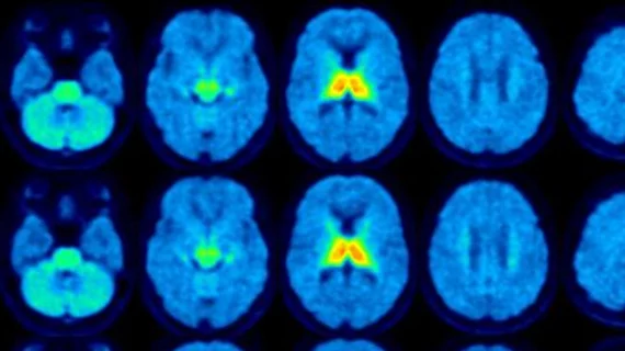
Molecular imaging (also called nuclear medicine or nuclear imaging) can image the function of cells inside the body at the molecular level. This includes the imaging modalities of positron emission computed tomography (PET) and single photon emission computed tomography (SPECT) imaging. How does PET and SPECT imaging work? Small amounts of radioactive material (radiopharmaceuticals) injected into a patient. These can use sugars or chemical traits to bond to specific cells. The radioactive material is taken up by cells that consume the sugars. The radiation emitted from inside the body is detected by photon detectors outside the body. Computers take the data to assemble images of the radiation emissions. Nuclear images may appear fuzzy or ghostly rather than the sharper resolution from MRI and CT.  But, it provides metabolic information at a cellular level, showing if there are defects in the function of the heart, areas of very high metabolic activity associated with cancer cells, or areas of inflammation, data not available from other modalities. These noninvasive imaging exams are used to diagnose cancer, heart disease, Alzheimer’s and Parkinson’s disease, bone disorders and other disorders. 

PET captures how social rejection triggers a feel-good reaction

Painful rejection and other social ills prompt the same opioid response in the brain as physical pain, according to new in vivo PET neuroimaigng of snubbed subjects. 

October 23, 2013

Society of Nuclear Medicine and Molecular Imaging now accepting abstracts

The Society of Nuclear Medicine and Molecular Imaging has begun accepting abstract submissions for the SNNMI 2014 Annual Meeting, held from June 7-11 in St. Louis.

October 22, 2013

Sofie Bio receives $1.8M SBIR grant for phase II clinical trials

Sofie Biosciences, an emerging in vivo imaging diagnostics company focused on PET probes, scanners and chemistry systems,announced today that the National Institute of Health has awarded the company a grant under the Small Business Innovation Research (SBIR) program.

October 2, 2013

Philips showcases innovations in radiation oncology imaging technology and workflow planning at the 55th American Society for Radiation Oncology (ASTRO) annual meeting

At the 55th American Society for Radiation Oncology ASTRO annual meeting, Sept. 22-25 in Atlanta, Royal Philips will feature the latest innovations that address efficient and personalized radiation oncology care for patients and their clinicians.

September 30, 2013

Siemens’ PETNET Solutions to provide PET radiopharmaceutical services for the US Oncology Network

Siemens’ PETNET Solutions has announced a three-year agreement with The US Oncology Network to supply its entire portfolio of Food and Drug Administration (FDA)-approved PET radiopharmaceutical agents at each of PETNET Solutions’ current Good Manufacturing Practices (cGMP)-certified locations throughout the United States.

September 19, 2013

Definiens and Advanced Cell Diagnostics launch software for quantitative RNA in situ hybridization

Definiens AG, a healthcare company that advances personalized medicine through image analysis and digital pathology solutions, and Advanced Cell Diagnostics (ACD) of Hayward California, a leader in molecular pathology, announced recently the commercial launch of RNAscope® SpotStudio™, a custom-designed image analysis software application for ACD's RNAscope®Assays to detect and quantify RNA biomarkers.

September 17, 2013

FDA clears Siemens’ Symbia Intevo integrated SPECT and CT system

Siemens Healthcare has announced that the FDA has granted 510(k) clearance for Symbia Intevo—the world’s first xSPECT system, which combines the high sensitivity of single-photon emission computed tomography (SPECT) with the high specificity of CT.

September 5, 2013

SNMMI 2013-2015 Wagner-Torizuka Fellowship Recipients Announced

The Society of Nuclear Medicine and Molecular Imaging (SNMMI) is pleased to announce the recipients of the 2013-2015 SNMMI Wagner-Torizuka Fellowship. This two-year fellowship, founded in 2008 by the late Henry N. Wagner, Jr., MD, and the late Kanji Torizuka, MD, PhD, is designed to provide extensive training and experience in the fields of nuclear medicine and molecular imaging for Japanese physicians in the early stages of their careers.

September 3, 2013

Around the web

Automated AI-generated measurements combined with annotated CT images can improve treatment planning and help referring physicians and patients better understand their disease, explained Sarah Jane Rinehart, MD, director of cardiac imaging with Charleston Area Medical Center.

Two advanced algorithms—one for CAC scores and another for segmenting cardiac chamber volumes—outperformed radiologists when assessing low-dose chest CT scans. 

"Gen AI can help tackle repetitive tasks and provide insights into massive datasets, saving valuable time," Thomas Kurian, CEO of Google Cloud, said Tuesday. 

Trimed Popup
Trimed Popup