Computed Tomography

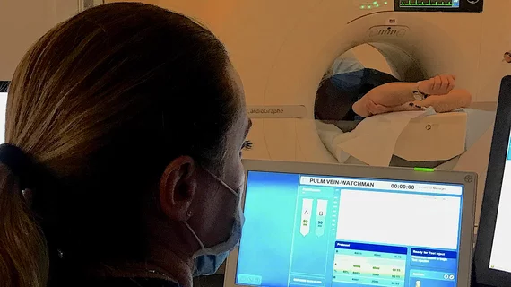
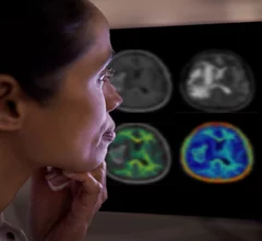
Computed tomography (CT) is a fast and accurate imaging modality often used in emergency settings and trauma imaging. CT scans, with or without (or both) iodinated contrast are frequently used to image the brain, chest, abdomen and pelvis, but also have post-imaging reconstructive capabilities for detailed orthopedic imaging. It is now a standard imaging modality in emergency rooms to quickly assess patients. CT uses a series of X-ray images shot as the gantry rotates around the patient. Computer technology assembles these into into a dataset volume than can be slices on any access, or advanced visualization software can extract specific parts of the anatomy for study. Find more content specific to cardiac CT.


john_constable

Scientists turn to medical imaging for deep analysis of John Constable painting

Researchers from the University of Bradford deployed CT, Raman spectroscopy, 3D microscopy and an X-ray to confirm the authenticity of one of Constable's landscapes.

April 1, 2024
fossil_evolution.jpg

Hospital, museum team up to CT scan dinosaur bones

The images will not only provide new scientific data, they will also serve as a digital backup of fragile fossils.

March 29, 2024

'Kitten Scanner' easing children's anxiety over CT imaging

The tiny Philips-branded CT unit isn't just a toy, it's an interactive simulation.

March 21, 2024
The rapid rise of artificial intelligence (AI) has helped cardiologists, radiologists, nurses and other healthcare providers embrace precision medicine in a way that ensures more heart patients are receiving personalized care.

FDA clears AI-enhanced 3D heart models generated from MR and CT images

The inHeart models significantly reduce cardiac ablation surgery times.

March 20, 2024
Head CT of a soldier with shrapnel wounds to head at a hospital in Dnipro. Photo by radiologist Olga Kachanova. Courtesy of RSNA.

PHOTO GALLERY: Radiology images of Ukrainian trauma patients wounded in war

A collection of medical images from hospitals in Ukraine showing radiology equipment in use and clinical images of wounded soldiers and civilians.


March 18, 2024
Micro-X mobile CT scanner

Mobile CT stroke detector passes VR simulation, set for field trials

The foldable unit weighs less than 154 pounds and can fit into an ambulance.

March 18, 2024
Radiologist using the Philips Smart Quant 3D Neuro artificial intelligence (AI) software to perform our measurements for white matter, gray matter and other parameters on brain MRI.

PHOTO GALLERY of brain imaging

This is a clinical photo gallery of neuro imaging and what conditions can be can be visualized in brain scans, and various imaging techniques used.

March 18, 2024

Outpatient study finds CT contrast is generally safe, but risk of adverse events varies with the seasons

A three-institution study of 473,482 patients found the risk of an adverse event following CT contrast increases in the summer and fall.

March 15, 2024

Around the web

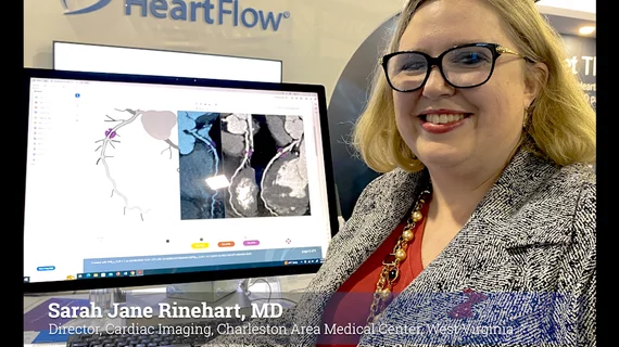
Automated AI-generated measurements combined with annotated CT images can improve treatment planning and help referring physicians and patients better understand their disease, explained Sarah Jane Rinehart, MD, director of cardiac imaging with Charleston Area Medical Center.

Two advanced algorithms—one for CAC scores and another for segmenting cardiac chamber volumes—outperformed radiologists when assessing low-dose chest CT scans. 

"Gen AI can help tackle repetitive tasks and provide insights into massive datasets, saving valuable time," Thomas Kurian, CEO of Google Cloud, said Tuesday. 

Trimed Popup
Trimed Popup