Computed Tomography

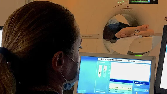
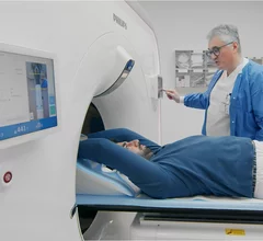
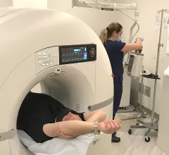
Computed tomography (CT) is a fast and accurate imaging modality often used in emergency settings and trauma imaging. CT scans, with or without (or both) iodinated contrast are frequently used to image the brain, chest, abdomen and pelvis, but also have post-imaging reconstructive capabilities for detailed orthopedic imaging. It is now a standard imaging modality in emergency rooms to quickly assess patients. CT uses a series of X-ray images shot as the gantry rotates around the patient. Computer technology assembles these into into a dataset volume than can be slices on any access, or advanced visualization software can extract specific parts of the anatomy for study. Find more content specific to cardiac CT.


Medical 3D printing expert Summer Decker, PhD, professor and vice chair for research and innovation, and director of 3D clinical applications, University of South Florida (USF) Morsani College of Medicine and Tampa General Hospital, explains what is involved in creating a 3D printiung program in radiology and how it helps patient care.

Pitching the business case for 3D printing labs in radiology

Medical 3D printing expert Summer Decker, PhD, University of South Florida, explains how her lab has helped improve patient care. 

March 14, 2024

New imaging protocols proposed to curb rise of cardiovascular infections

Eleven medical societies have signed on to a consensus statement aimed at standardizing imaging for suspected cardiovascular infections.

March 12, 2024
An MRI of a fetus inside the mother. The freakish appearance of the eyes and the face are normal for MRI fetal imaging. Image courtesy of RSNA. Baby MRI, baby ultrasound

PHOTO GALLERY: What does fetal medical imaging look like?

This is a clinical photo gallery of fetal imaging that explains what all can be seen on medical imaging, how sex is determined, how measurements are used to track the development of a baby.

March 11, 2024
Fish

Museum project uses contrast CT and X-ray to catalog world’s vertebrates

Over the course of five years, 18 different institutions worked to produce the images for a Florida Museum initiative.

March 6, 2024
Using computed tomography (CT) to perform coronary artery calcium (CAC) scoring can help identify symptomatic chest pain patients who do not require further testing, according to a new analysis published in Radiology.[1]

Coronary calcium scoring predicts when chest pain patients can skip invasive testing

Using CT to perform coronary artery calcium scoring on symptomatic chest pain patients can deliver significant value, according to a new data published in Radiology

March 6, 2024
Kate Hanneman, MD, University Of Toronto, explains why vendors and hospitals are increasingly discussing lowing their carbon footprint by starting with radiology. 

What does radiology have to do with climate change?

Kate Hanneman, MD, explains why many vendors and hospitals want to lower radiology's impact on the environment. "Taking steps to reduce the carbon footprint in healthcare isn’t just an opportunity," she said. "It’s also a responsibility."

March 1, 2024
The new Philips Healthcare CT 5300 system is aimed at the cardiac CT market and incorporates AI features to improve image quality and workflow. #ECR #YesCCT #CCTA

Philips launches new AI-enabled CT scanner aimed at cardiology at ECR 2024

Philips introduced a new CT system at ECR aimed at the rapidly growing cardiac CT market, incorporating numerous AI features to optimize workflow and image quality.

March 1, 2024
A medical professional trained to manage contrast reactions should be on-site at any imaging facilities doing contrast studies to maintain patient safety, according to a an updated statement from the American College of Radiology. The ACR has released the latest edition of the ACR Appropriateness Criteria, which includes 221 diagnostic imaging and interventional radiology topics with more than 1,050 clinical variants covering 2,900 clinical scenarios. #ACR #AUC #appropriateuse

ACR updates requirements for on-site staff overseeing contrast administration

A clinician trained to manage reactions should be on-site at any imaging facilities doing contrast studies to maintain patient safety.

March 1, 2024

Around the web

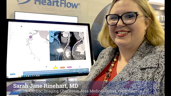
Automated AI-generated measurements combined with annotated CT images can improve treatment planning and help referring physicians and patients better understand their disease, explained Sarah Jane Rinehart, MD, director of cardiac imaging with Charleston Area Medical Center.

Two advanced algorithms—one for CAC scores and another for segmenting cardiac chamber volumes—outperformed radiologists when assessing low-dose chest CT scans. 

"Gen AI can help tackle repetitive tasks and provide insights into massive datasets, saving valuable time," Thomas Kurian, CEO of Google Cloud, said Tuesday. 

Trimed Popup
Trimed Popup