Molecular Imaging

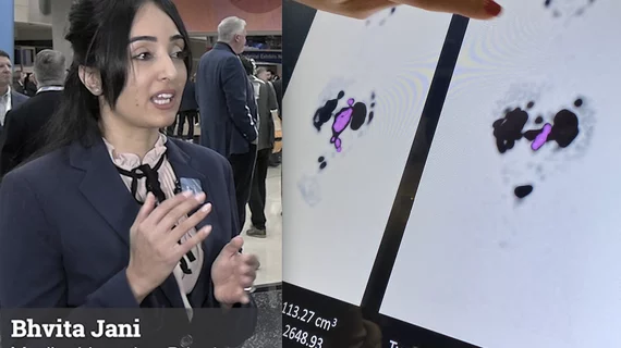
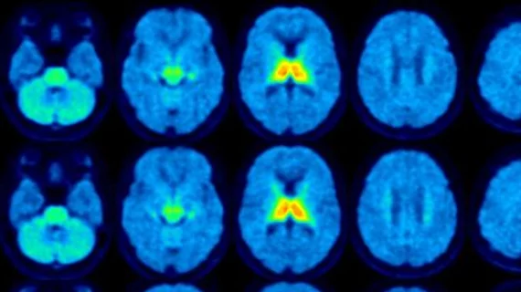
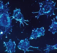
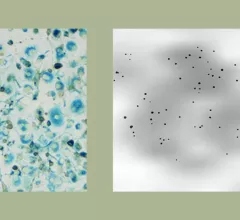
Molecular imaging (also called nuclear medicine or nuclear imaging) can image the function of cells inside the body at the molecular level. This includes the imaging modalities of positron emission computed tomography (PET) and single photon emission computed tomography (SPECT) imaging. How does PET and SPECT imaging work? Small amounts of radioactive material (radiopharmaceuticals) injected into a patient. These can use sugars or chemical traits to bond to specific cells. The radioactive material is taken up by cells that consume the sugars. The radiation emitted from inside the body is detected by photon detectors outside the body. Computers take the data to assemble images of the radiation emissions. Nuclear images may appear fuzzy or ghostly rather than the sharper resolution from MRI and CT.  But, it provides metabolic information at a cellular level, showing if there are defects in the function of the heart, areas of very high metabolic activity associated with cancer cells, or areas of inflammation, data not available from other modalities. These noninvasive imaging exams are used to diagnose cancer, heart disease, Alzheimer’s and Parkinson’s disease, bone disorders and other disorders. 

australian-flag-map.jpg

Australian reactor to begin producing Tc-99m following shutdown

Production of technetium-99m (Tc-99m) is expected to resume soon at the Australian generator that was forced to shut down in June, according to the Australian Nuclear Science and Technology Organization (ANSTO).

August 9, 2018

PET imaging method could improve Type 1 diabetes treatment

Yale University researchers have discovered a new PET imaging method that measures beta-cell mass which could improve monitoring, according to research published online in the August issue of the Journal of Nuclear Medicine.  

August 7, 2018

PET tracer IDs estrogen receptor variation in breast cancer patients

Dutch researchers utilized a PET tracer to distinguish differences in estrogen receptor (ER) expression in metastatic breast cancer patients, according to a recent Journal of Nuclear Medicine study. Findings may enhance treatment for these patients.

August 7, 2018

Molecular imaging tool can map cancer cell division in real-time

Researchers from Columbia University in New York have developed a molecular imaging tool that can track metabolic changes in individual living cells in real time, according to research published July 30 in Nature Communications.

August 6, 2018
mri-resolution-stevens-web-824x549.jpg

MRI enhances cellular resolution to determine effectiveness of cancer therapy

Researchers from the Keck School of Medicine at the University of Southern California (USC) successfully used high-resolution MRI and a cell-labeling technique to visualize nearly 100 cells, which may allow clinicians to assess the effectiveness of immune cell- and stem cell-based therapies to treat cancer.

July 25, 2018

Can a new clinical state for prostate cancer help personalize treatment for the disease?

For patients with metastatic prostate cancer, understanding its genesis and evolution can mean all the difference for treatment. But, what if, as a recent Journal of Nuclear Medicine perspective proposes, a new “clinical state” could improve understanding and treatment of the disease?

July 23, 2018
SNMMI

SNMMI, imaging leaders back $20M in federal funds for Mo-99 production

The Society for Nuclear Medicine and Molecular Imaging (SNMMI), along with a host of other leading medical imaging groups and vendors, addressed a July 18 letter to the Senate and House subcommittees on Energy and Water Development voicing its support for $20 million in additional funding to help establish domestic production of molybdenum-99 (Mo-99).

July 23, 2018

PET imaging helps map Parkinson’s damage to heart

A team from the University of Wisconsin-Madison successfully used PET imaging to visualize heart damage commonly seen in those with Parkinson’s disease. The technique will help develop therapies.

July 16, 2018

Around the web

Automated AI-generated measurements combined with annotated CT images can improve treatment planning and help referring physicians and patients better understand their disease, explained Sarah Jane Rinehart, MD, director of cardiac imaging with Charleston Area Medical Center.

Two advanced algorithms—one for CAC scores and another for segmenting cardiac chamber volumes—outperformed radiologists when assessing low-dose chest CT scans. 

"Gen AI can help tackle repetitive tasks and provide insights into massive datasets, saving valuable time," Thomas Kurian, CEO of Google Cloud, said Tuesday. 

Trimed Popup
Trimed Popup