Molecular Imaging

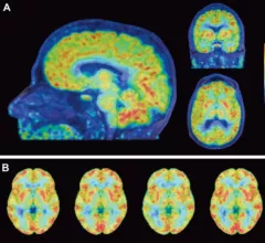
Molecular imaging (also called nuclear medicine or nuclear imaging) can image the function of cells inside the body at the molecular level. This includes the imaging modalities of positron emission computed tomography (PET) and single photon emission computed tomography (SPECT) imaging. How does PET and SPECT imaging work? Small amounts of radioactive material (radiopharmaceuticals) injected into a patient. These can use sugars or chemical traits to bond to specific cells. The radioactive material is taken up by cells that consume the sugars. The radiation emitted from inside the body is detected by photon detectors outside the body. Computers take the data to assemble images of the radiation emissions. Nuclear images may appear fuzzy or ghostly rather than the sharper resolution from MRI and CT.  But, it provides metabolic information at a cellular level, showing if there are defects in the function of the heart, areas of very high metabolic activity associated with cancer cells, or areas of inflammation, data not available from other modalities. These noninvasive imaging exams are used to diagnose cancer, heart disease, Alzheimer’s and Parkinson’s disease, bone disorders and other disorders. 

PET/CT diagnoses blood vessel disease better than biopsy method

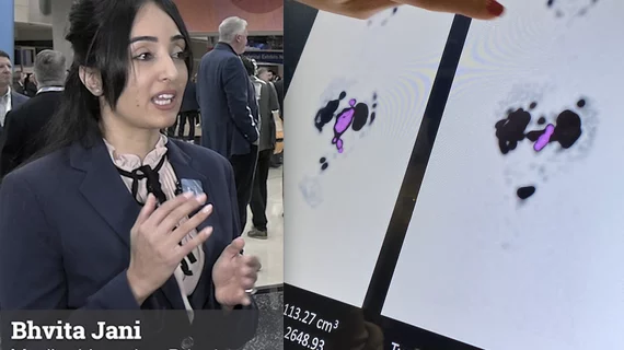
Pairing PET with CT achieved better diagnostic accuracy than temporal artery biopsy (TAB) in patients with newly suspected giant cell arteritis (GCA), according to research presented recently at the 2018 American College of Rheumatology (ACR)/ Association of Reproductive Health Professional (AHRP) meeting in Chicago.

October 22, 2018
screen_shot_2018-10-22_at_10.03.42_am.png

Novel molecular CT technique could revolutionize drug discovery

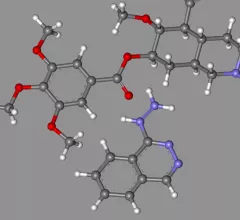
The technique is able to map the structure of small organic molecules, such as pharmaceuticals and hormones, with electron diffraction imaging—commonly used to chart larger proteins, according to a report published Oct. 19 by Science Magazine.

October 22, 2018

PET quantification method can help improve amyloid pathology assessment

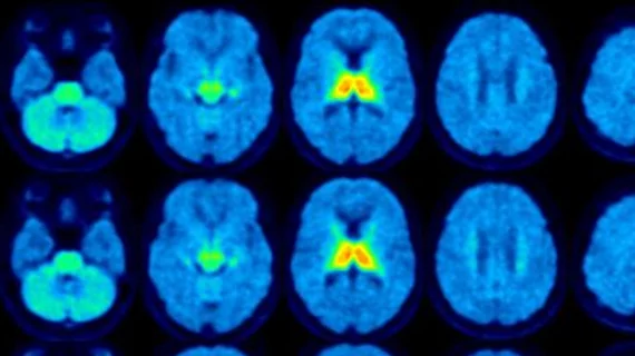
Utilizing non-displaceable binding potential (BPND) quantification with 18F-Flutemetamol PET imaging can better assess amyloid pathology in elderly patients with low amyloid buildup compared to traditional standard uptake value ratio (SUVr), reported authors of an Oct. 12 study in the Journal of Nuclear Medicine.

October 12, 2018

PET can image T-cells for chronic liver disease

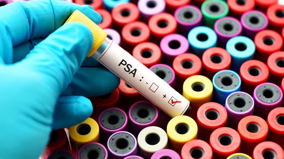
PET imaging could serve as an accurate, noninvasive substitute to liver biopsies in patients with chronic liver diseases, as detailed in research published in the October issue of The Journal of Nuclear Medicine.

October 10, 2018

Molecular imaging research could find less toxic, more effective breast cancer treatments

Researchers from the Medical University of South Carolina (MUSC) Hollings Cancer Center in Charleston have received a three-year, $1.2 million grant from the U.S. Department of Defense to use molecular imaging tools to test antibody therapies for breast cancer—potentially producing fewer side effects.

October 8, 2018
Price transparency study in radiology

NorthStar may receive federal funding to produce Mo-99

NorthStar Medical Radioisotopes in Beloit, Wis. and the Janesville, Wis.-planned SHINE Medical Technologies may receive a portion of the $20 million in federal technology funding bill approved by President Donald Trump to produce molybdenum-99 (Mo-99).

October 8, 2018

Nanoplatform developed with 3 molecular imaging modalities may improve cancer diagnosis

A new hybrid nanoplatform that simultaneously uses three types of imaging modalities—MRI, CT and fluorescence optical imaging—to locate tumors could give a new edge to molecular imaging and tumor diagnosis.  

October 5, 2018
gi_66154_po1a0230_gold-1500x1000.png

Cancer imaging researcher receives highest honor from World Molecular Imaging Society

The World Molecular Imaging Society (WMIS) has awarded Robert Gillies, PhD, the 2018 Gold Medal Award to honor his lifetime of research in cancer molecular imaging and cancer biology, according to a WMIS press release.

September 27, 2018

Around the web

Two advanced algorithms—one for CAC scores and another for segmenting cardiac chamber volumes—outperformed radiologists when assessing low-dose chest CT scans. 

"Gen AI can help tackle repetitive tasks and provide insights into massive datasets, saving valuable time," Thomas Kurian, CEO of Google Cloud, said Tuesday. 

SCAI and four other major healthcare organizations signed a joint letter in support of intravascular ultrasound. 

Trimed Popup
Trimed Popup